
Why a Neck Disc Prolapse Needs Different Surgery Than the Back
Is anterior cervical disc surgery more dangerous than lumbar disc surgery?
It involves navigating more complex anatomy, including major blood vessels and airway structures, and therefore requires specific surgical training and experience. However, when performed by an appropriately trained and experienced surgeon, it is a very well-established procedure with a strong track record of safety and good outcomes.
Will I lose movement in my neck after this surgery?
A single-level anterior cervical discectomy and fusion typically results in a relatively small reduction in overall neck mobility, as the fused level no longer moves independently, though most patients adapt well and retain good functional range of motion from the remaining mobile segments. Multi-level fusions result in a greater reduction in mobility.
How long does recovery take after ACDF surgery?
Most patients are able to go home within one to two days of surgery. Return to light activities typically takes two to four weeks, with full fusion and consolidation of the bone graft taking approximately three to six months, though most patients feel substantially improved well before this point.
Can I have a sore throat or difficulty swallowing after this surgery?
Mild and temporary difficulty swallowing or a sore throat is common in the days following anterior cervical surgery, related to the gentle retraction of the oesophagus and surrounding soft tissues during the procedure. This typically resolves within a few days to a couple of weeks as the local tissue swelling settles.
What is the difference between a cage and a plate in cervical disc surgery?
The cage is the structural spacer device, filled with bone graft, that is placed directly within the disc space to restore height and provide a scaffold for fusion. A plate, when used, is an additional thin metal piece placed across the front of the vertebrae and secured with screws to provide further immediate stability to the construct while the fusion consolidates.
Is cervical disc replacement a better option than fusion?
Neither option is universally better. Disc replacement preserves motion at the treated level and may reduce stress on adjacent segments over time, making it an attractive option for some younger patients with single-level disease. Fusion provides definitive, proven stability and remains the appropriate choice in many clinical situations. The decision is individualised based on the patient’s specific anatomy and circumstances.
A disc prolapse in the lower back and a disc prolapse in the neck are, at the cellular and structural level, fundamentally the same kind of problem: disc material pushing against and compressing a nearby nerve. Yet the surgical approach used to treat them is completely different, dictated entirely by a single anatomical reality that has nothing to do with the disc itself and everything to do with what sits directly behind it.
Dr. Sherief Elsayed, Consultant Spine Surgeon in Dubai, explains exactly why cervical disc surgery cannot simply mirror lumbar disc surgery, and walks through what the alternative approach actually involves.
The Straightforward Case: Lumbar Disc Surgery
Dr. Sherief Elsayed begins by describing the comparatively simple lower back procedure: “If you have a disc prolapse in your lower back, it’s usually pretty straightforward for surgery. We come in, we move the nerves out of the way, take out the disc prolapse, nerves flop back, everything’s all right.”
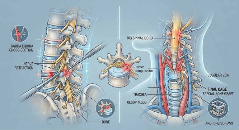
This description captures the essential simplicity of a standard posterior lumbar microdiscectomy. The surgeon approaches directly from the back of the spine. Because, at this lumbar level, the structure within the spinal canal consists of individual nerve roots, the cauda equina, rather than the spinal cord itself, these nerve roots can be safely and gently retracted to one side, creating a working space through which the surgeon can remove the herniated disc material. Once removed, the nerve root returns to its normal position, no longer compressed, and the procedure is essentially complete from a decompression standpoint.
The Anatomical Obstacle in the Neck
The same straightforward posterior approach simply cannot be used in the cervical spine, and Dr. Sherief Elsayed identifies the precise reason why: “The reason that we can’t come from the back is because there’s a big spinal cord in the way. We can’t move that out of the way.”
This is the critical anatomical distinction between the lumbar and cervical regions of the spine. The spinal cord itself, the central neurological structure connecting the brain to the rest of the body via the highway described in the published article Why Spinal Cord Injuries Cause Different Symptoms, A UAE Spine Surgeon Explains, occupies the spinal canal throughout the cervical region. Unlike the individual, more mobile nerve roots of the cauda equina in the lower lumbar spine, the spinal cord cannot be safely retracted or moved aside even briefly. Any significant manipulation or compression of the cord risks causing immediate and potentially permanent neurological injury, including paralysis.
This single anatomical fact, the presence of the spinal cord directly behind the cervical discs, is what makes the posterior surgical approach used so routinely in the lower back simply unsafe to apply in the same way to the neck.
The Solution: Approaching From the Front
Because the spinal cord makes a direct posterior approach to the disc unsafe in the cervical spine, surgeons instead approach from the opposite direction entirely. Dr. Sherief Elsayed explains: “If on the other hand you have a disc prolapse up in your neck, more often than not, we need to come in from the front.”
This describes an anterior cervical approach, one of the most commonly performed and well-established procedures in all of spinal surgery.
The anatomical structures navigated during this approach:
Dr. Sherief Elsayed details the specific anatomy that must be carefully identified and protected during this approach: “Carotid artery, jugular vein to one side, trachea and oesophagus to the other side, and then down onto the spine.”
This description reflects the genuine anatomical complexity of the front of the neck. The carotid artery, supplying blood to the brain, and the internal jugular vein, draining blood from the head, run together in a protected sheath on one side of the surgical corridor. The trachea (windpipe) and oesophagus (the tube carrying food to the stomach) sit in the midline structures that must be gently retracted, rather than the major vessels, to create the surgical pathway. By working carefully between these structures, all of which must be identified, protected, and in the case of the trachea and oesophagus, gently moved aside rather than cut or damaged, the surgeon reaches the front surface of the cervical spine directly, with the spinal cord remaining safely undisturbed behind the vertebral bodies and discs throughout the entire approach.
This anterior corridor is, in many respects, a more anatomically complex pathway to navigate. A Thoracic Spine Surgeon in Dubai trained across the full spinal column brings this same careful anatomical approach to procedures throughout the spine, recognising the additional complexity compared to the posterior approach used in the lumbar spine. However, it offers the decisive safety advantage of reaching the cervical disc without ever needing to manipulate or retract the spinal cord itself.
What Happens Once the Disc Is Reached
Having safely navigated to the front of the affected cervical level, the surgical removal and reconstruction process follows a specific sequence.
Dr. Sherief Elsayed describes the steps: “When we come in from the front, we take out the disc. We use a trial in its place in order to get the right size, and then we insert our final cage. That cage is either held by some anchors or by screws, and inside the cage we put special bone graft in which helps the bone to fuse together.”
The surgical sequence in detail:
- Disc removal: The herniated and remaining disc material at the affected level is carefully removed, directly decompressing the nerve root or spinal cord that was being compressed from behind by the original prolapse
- Trial sizing: A trial spacer, a sizing instrument, is placed temporarily into the now-empty disc space to determine the precise height and dimensions required for the final implant, ensuring an accurate fit that restores appropriate disc height without over-distracting the surrounding soft tissues
- Cage insertion: Once the correct size has been confirmed using the trial, the final cage, a structural implant designed to occupy the disc space permanently, is inserted
- Securing the cage: The cage is secured in position using either small integrated anchors that grip directly into the adjacent vertebral bone, or separate screws, sometimes combined with a small anterior plate, providing immediate stability to the construct
- Bone graft for fusion: Bone graft material is packed within the hollow centre of the cage itself, providing the biological material necessary to stimulate new bone growth and ultimately achieve solid fusion between the two adjacent vertebrae over the following months
This procedure is known as Anterior Cervical Discectomy and Fusion, commonly abbreviated as ACDF, and represents one of the most extensively studied and reliably effective procedures in spinal surgery, with a long track record of safety and good clinical outcomes for appropriately selected patients.
Why Fusion Is Typically Required in the Cervical Approach
A notable difference between the lumbar and cervical procedures described here is that the lumbar microdiscectomy, as Dr. Sherief Elsayed describes, generally does not require fusion. The nerve is decompressed, and the disc, though it has had material removed, remains otherwise in place.
In the anterior cervical approach, by contrast, because the entire disc is typically removed during the procedure to safely access and decompress the underlying neural structures, something must be placed in the resulting empty disc space both to restore appropriate disc height and to provide structural support for the spine. The cage filled with bone graft serves this purpose, and the resulting fusion provides long-term stability to the operated level once healing is complete.
For carefully selected patients, particularly younger individuals at a single level without significant adjacent degeneration, an alternative to fusion exists. A Spine Fusion Surgeon in Dubai can advise whether disc replacement or fusion is the more appropriate choice for your specific anatomy: cervical disc replacement uses a mobile prosthetic implant designed to preserve some motion at the treated level rather than fusing it. The choice between fusion and disc replacement is made individually based on the patient’s specific anatomy, age, and the clinical situation.
Why This Difference Matters for Patients
Patients who have heard about a friend or family member’s relatively straightforward lumbar disc surgery sometimes expect a similarly simple recovery and procedure if they themselves are diagnosed with a cervical disc prolapse. Understanding that the cervical procedure is anatomically more involved, requiring careful navigation around major blood vessels and airway structures, and typically involves permanent implantation of a cage and bone graft material, helps set appropriate and accurate expectations for both the surgery itself and the recovery process that follows.
A Cervical Spine Doctor in Dubai will explain this specific anatomical rationale clearly to any patient being considered for cervical disc surgery, ensuring the decision to proceed is made with a full and accurate understanding of what the procedure actually involves and why it differs so significantly from lumbar disc surgery.
UAE-Specific Considerations
Anterior cervical discectomy and fusion is a well-established and commonly performed procedure at specialist spinal surgical centres throughout Dubai and the wider UAE, performed by surgeons with appropriate fellowship training in cervical spine surgery. Patients diagnosed with a cervical disc prolapse causing arm pain, numbness, or weakness should seek assessment with a surgeon experienced specifically in this anterior approach, given its anatomical complexity relative to the more routine posterior lumbar procedures.
Expert Summary
The presence of the spinal cord directly behind the cervical discs, a structure that simply cannot be safely retracted or moved aside the way the more mobile lumbar nerve roots can, is the single anatomical fact that necessitates an entirely different surgical strategy for neck disc prolapse compared to lower back disc prolapse. Approaching from the front, carefully navigating between the major blood vessels and airway structures of the neck, allows the surgeon to reach and remove the cervical disc while leaving the spinal cord completely undisturbed throughout the procedure. The subsequent insertion of a cage filled with bone graft, secured with anchors or screws, both restores disc height and achieves the fusion needed for long-term stability at the treated level.
Table of Contents
Recent Articles
Why Eight People Were Needed for One Spine Surgery in Dubai
Why Eight People Were Needed for One Spine Surgery in Dubai Does every spinal surgery require exactly eight team members? No, as Dr. Sherief Elsayed explains directly, the specific team
Three Spine Surgery Myths a Dubai Surgeon Wants You to Forget
Three Spine Surgery Myths a Dubai Surgeon Wants You to Forget Is it really safe to walk on the same day as spine surgery? Yes, for the majority of modern
Why Osteoporosis Is Called the Silent Disease in the UAE
Why Osteoporosis Is Called the Silent Disease in the UAE If osteoporosis doesn’t cause pain, how would I know if I needed a DEXA scan? Given the entirely silent nature