
How Nerve Pain Patterns Help Diagnose Sciatica in the UAE
Can dermatomal pattern alone diagnose the exact cause of my nerve pain?
Dermatomal pattern strongly suggests which specific nerve root is likely involved, but it does not by itself identify the underlying structural cause, whether that is a disc herniation, spinal stenosis, or another condition. Imaging is needed to confirm the specific cause once the likely nerve level has been identified through clinical assessment.
Why do some patients have pain that doesn't clearly follow a textbook dermatomal pattern?
Individual anatomical variation in nerve root distribution is common, and some patients have overlapping innervation from adjacent levels that can blur the classic pattern. Additionally, some pain sources are not nerve root-related at all, which is why the full clinical assessment considers multiple possibilities rather than relying on dermatomal pattern alone.
Is dermatomal testing painful or uncomfortable?
No, dermatomal assessment is generally performed through gentle, non-invasive testing of light touch or pinprick sensation across different areas of skin, compared between the affected and unaffected sides of the body, and causes no significant discomfort.
Can dermatomal changes be permanent?
This depends on the underlying cause and how promptly and effectively it is treated. As discussed in the earlier article on nerve compression timing, prompt treatment of significant nerve compression generally offers the best chance of full sensory recovery, though some degree of residual change can persist in certain cases, particularly with prolonged or severe compression.
Do myotomes work the same way as dermatomes?
Yes, myotomes follow a similar principle but apply to muscle groups rather than skin sensation, with each spinal nerve root supplying a specific, mapped set of muscles. Testing the strength of these muscle groups, alongside dermatomal sensory testing, provides complementary information supporting the same overall diagnostic process.
Should I describe my pain pattern in detail to my doctor even if it seems obvious?
Yes, the precise distribution of radiating pain, numbness, or tingling is genuinely valuable diagnostic information. Describing exactly where the pain starts, where it travels to, and any associated numbness or tingling in specific fingers or toes provides your treating specialist with important clinical detail that directly informs their assessment.
Before a single image has been taken, an experienced spine specialist can already form a strong clinical impression of which specific nerve is likely affected in a patient with radiating pain, based entirely on precisely where that pain travels in the body. This is not guesswork. It reflects a detailed anatomical map, developed and refined over more than a century of clinical neurology, that links specific areas of skin sensation to specific spinal nerve roots.
Dr. Sherief Elsayed, Consultant Spine Surgeon in Dubai, explains this diagnostic principle directly, using specific clinical examples that demonstrate exactly how this pattern recognition works in practice.
What Are Dermatomes?
Dr. Sherief Elsayed introduces the concept clearly: “Dermatomes are like a map of the nerves and which part of the body they supply in terms of skin sensation.”
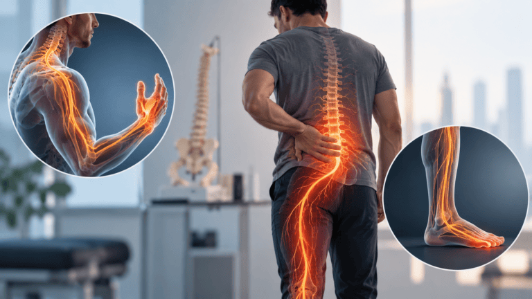
A dermatome is a specific area of skin that is primarily supplied by sensory fibres from a single spinal nerve root. As spinal nerve roots exit the spinal cord and travel outward to supply their target areas, each one innervates a reasonably predictable and mapped region of skin. This means that when a specific spinal nerve root is irritated or compressed, whether from a disc herniation, spinal stenosis, or another cause, the resulting pain, numbness, or tingling characteristically follows the specific dermatomal pattern associated with that particular nerve root, rather than appearing in a random or unpredictable distribution.
This dermatomal map has been established through more than a century of careful clinical observation and neuroanatomical study, and it remains one of the most fundamental and clinically useful tools in the assessment of any patient presenting with radiating nerve pain.
Clinical Example One: Neck and Arm Pain Into the Thumb
Dr. Sherief Elsayed provides a specific clinical scenario: “If I have a patient that presents with severe neck pain, arm pain, going down into his thumb, I’m thinking C6 dermatome.”
The C6 nerve root, exiting the cervical spine between the sixth and seventh cervical vertebrae, has a well-established dermatomal distribution that runs down the lateral (outer) aspect of the arm and forearm, terminating in the thumb and index finger. A patient describing pain that specifically radiates from the neck, down the outer arm, and into the thumb is describing a pattern that matches this C6 distribution precisely.
This single piece of history, described in the patient’s own words before any physical examination or imaging has taken place, already generates a specific, testable clinical hypothesis: compression or irritation of the C6 nerve root, most commonly from a cervical disc herniation or degenerative changes at the C5/C6 level, given that spinal nerve roots typically exit above their correspondingly numbered vertebra in the cervical spine.
Clinical Example Two: Sciatica Into the Outside of the Foot
Dr. Sherief Elsayed provides a corresponding example from the lumbar spine: “If the patient has sciatica and it’s going down into the outside of his foot, I’m thinking the S1 dermatome.”
The S1 nerve root has a dermatomal distribution that runs down the back of the thigh and calf, terminating along the outside (lateral) border of the foot and the small toe. A patient describing classic sciatica, meaning pain radiating from the lower back through the buttock and down the leg, specifically travelling to the outer aspect of the foot, is describing a pattern consistent with S1 nerve root involvement, most commonly from an L5/S1 disc herniation.
Why This Matters: Predicting the Imaging Before Seeing It
Dr. Sherief Elsayed makes the clinical significance of this pattern recognition explicit: “So before I’ve even seen the MRI, I have an idea of what the MRI scan is going to show.”
This is a genuinely important demonstration of how good clinical reasoning works in spine medicine, and it connects directly to a theme that has recurred throughout this blog series: the relationship between clinical examination and imaging findings.
Why this predictive capacity matters clinically:
As established repeatedly throughout earlier articles in this series, including the discussion in When Is Spine Surgery Wrong? Dr. Sherief Elsayed Explains, the value of an imaging finding depends entirely on whether it correlates with the patient’s actual clinical presentation. Dermatomal pattern recognition provides exactly this correlation check, in advance.
When a patient describes a C6 distribution and the subsequent MRI confirms a C5/C6 disc herniation compressing the C6 nerve root, this alignment between history, examination, and imaging provides strong confidence that the identified structural finding is genuinely the source of the patient’s symptoms, precisely the kind of correlation that supports appropriate, targeted treatment.
Conversely, if a patient describes a clear C6 dermatomal pattern but the MRI shows only mild changes at an entirely different level, with no significant pathology at C5/C6 specifically, this mismatch is itself clinically important information, prompting further consideration of whether an alternative diagnosis, a different affected level than initially suspected, or even a non-spinal cause of the symptoms might be present.
The Broader Dermatomal Map
While Dr. Sherief Elsayed’s two examples illustrate the principle clearly, the complete dermatomal map extends across the entire spine, with each level having its own characteristic distribution.
Common cervical dermatomes:
C5 supplies the lateral aspect of the upper arm and shoulder. C6, as described, extends to the thumb and index finger. C7 extends to the middle finger. C8 extends to the ring and little fingers and the medial (inner) aspect of the forearm.
Common lumbar and sacral dermatomes:
L4 supplies the medial aspect of the lower leg and the inner ankle. L5 extends to the top of the foot and the big toe. S1, as described, extends to the outer foot and little toe.
Each of these patterns, when described accurately by a patient and correlated with specific examination findings including reflexes, muscle strength testing in the corresponding myotome (the muscle group supplied by that same nerve root), and provocative testing, builds a comprehensive clinical picture that guides both the interpretation of subsequent imaging and the overall diagnostic confidence before any structural investigation has even been performed.
Why Dermatomal Assessment Complements Rather Than Replaces Imaging
It is important to note that dermatomal pattern recognition, however clinically valuable, is a component of the overall diagnostic process rather than a replacement for imaging entirely. Individual anatomical variation means that dermatomal boundaries are not always perfectly sharp or identical between every patient, some patients have overlapping innervation from adjacent nerve roots that can blur the classic textbook pattern, and imaging remains essential for confirming the specific structural cause, ruling out alternative or additional pathology, and providing the detailed anatomical information needed for surgical planning where relevant. A Spine Consultant in Dubai uses this alongside imaging to build the most accurate overall clinical picture.
A Nerve Compression Specialist in Dubai uses dermatomal history and examination findings as one important component of a comprehensive assessment, working alongside imaging rather than as a substitute for it, to build the most accurate and reliable overall clinical picture.
Why This Skill Matters for Patient Trust and Efficiency
There is a genuine practical benefit for patients when a specialist demonstrates this kind of clinical reasoning openly. A patient who is told, based on their described symptoms alone and before any scan has been reviewed, exactly what pattern of findings the treating specialist expects to see, and then has that expectation confirmed by the subsequent imaging, gains confidence that their assessment has been thorough and that the eventual treatment plan is genuinely grounded in an accurate, well-correlated diagnosis.
This approach also has practical efficiency benefits, helping to direct which specific spinal region and levels warrant the most detailed imaging attention, and providing an important cross-check against imaging findings that might otherwise be over-interpreted or under-interpreted in isolation, a theme explored extensively in the earlier article Why an MRI Report Alone Is Not Enough to Decide on Spine Treatment in the UAE.
UAE-Specific Context
Given the significant burden of both cervical and lumbar disc-related nerve compression seen across the UAE’s patient population, discussed extensively throughout this blog series, dermatomal history-taking is a routine and important part of the standard clinical assessment applied by spine specialists across Dubai. Patients presenting with radiating arm or leg pain should expect their treating specialist to ask detailed questions about the precise distribution of their symptoms, recognising that these details carry genuine diagnostic weight beyond simply confirming “there is pain.”
Expert Summary
Dermatomes provide a detailed anatomical map linking specific areas of skin sensation to specific spinal nerve roots, allowing an experienced clinician to form a strong, testable clinical impression of which nerve root is likely affected based entirely on the precise pattern of a patient’s described pain, numbness, or tingling. Dr. Sherief Elsayed’s examples, C6 pain travelling to the thumb and S1 sciatica travelling to the outer foot, demonstrate how this pattern recognition allows a clinician to predict imaging findings before they are seen, and provides an essential correlation check that strengthens diagnostic confidence when history, examination, and imaging findings align.
Table of Contents
Recent Articles

Why Eight People Were Needed for One Spine Surgery in Dubai
Why Eight People Were Needed for One Spine Surgery in Dubai Does every spinal surgery require exactly eight team members? No, as Dr. Sherief Elsayed explains directly, the specific team

Three Spine Surgery Myths a Dubai Surgeon Wants You to Forget
Three Spine Surgery Myths a Dubai Surgeon Wants You to Forget Is it really safe to walk on the same day as spine surgery? Yes, for the majority of modern
Why Osteoporosis Is Called the Silent Disease in the UAE
Why Osteoporosis Is Called the Silent Disease in the UAE If osteoporosis doesn’t cause pain, how would I know if I needed a DEXA scan? Given the entirely silent nature