
Why Lung Cancer Can Spread to the Spine, Dr. Sherief Elsayed Explains
Can back pain be the first sign of cancer spread to the spine?
Yes. New back pain in a patient with known cancer should be investigated for spinal metastasis. Even in patients without a known cancer diagnosis, persistent unexplained back pain that is worse at night, associated with weight loss, or accompanied by systemic symptoms warrants investigation for malignancy.
How quickly can MSCC develop?
MSCC can develop over hours, as in this case, or gradually over days to weeks depending on the mechanism. Rapid progression, as seen with vertebral collapse, is the most time-critical scenario. Any new or rapidly worsening neurological symptoms in a patient with cancer require emergency assessment.
What is the difference between a lumbar and thoracic MRI?
They image different sections of the spine. A lumbar MRI typically covers L1 to S1. A thoracic MRI covers T1 to T12. The clinical findings determine which region to image. When upper motor neurone signs are present, the lesion is at or above the thoracic cord and a lumbar MRI will not show it.
Can lung cancer in the spine be cured?
Spinal metastases from lung cancer are generally treated with palliative intent. The goal is to preserve neurological function, relieve pain, and maintain quality of life rather than achieve cure. Some patients with oligometastatic disease (limited metastases) treated with aggressive local therapy may achieve prolonged remission.
What should I do if I have cancer and develop back pain?
Contact your oncology team immediately and ask for urgent spinal imaging. Do not wait to see if the pain resolves. A spine MRI covering the symptomatic level is the most appropriate investigation. If neurological symptoms are also present, this is a medical emergency requiring same-day assessment.
Is surgery always necessary for metastatic spinal cord compression?
Not always. Patients with radiosensitive tumours (such as lymphoma or myeloma) and without significant spinal instability may be treated with radiotherapy alone. Patients with structural instability or rapidly progressing neurological deficit are best served by surgical decompression and stabilisation followed by radiotherapy.
A 45-year-old man who cannot walk. Bilateral leg weakness developing over hours. Severe thoracic back pain. A history of lung cancer diagnosed one year earlier. These details, assembled in the right sequence, point to one of the most time-critical diagnoses in spine medicine: metastatic spinal cord compression.
Dr. Sherief Elsayed, Consultant Spine Surgeon in Dubai, uses a case presentation approach to demonstrate how rapidly this diagnosis should be reached, what investigation is needed without delay, and why a lumbar MRI, however quickly obtained, can be the wrong scan in the wrong place.
The Clinical Case: 30 Seconds to Diagnosis
The presentation unfolds as a rapid clinical exchange:
“45 year old male walks in with weakness in both legs. How quickly did his weakness develop? Over a few hours. He’s completely unable to walk. Does he have any back pain? He has severe thoracic back pain. Has he had an MRI scan? He has had an MRI of his lumbar spine. It doesn’t show cauda equina syndrome. Clinical examination? Power is two out of five in all lower limb myotomes, up-going plantars, pathological clonus and hyperreflexia. Any history of malignancy? He had lung cancer diagnosed a year ago. My best guess would be a metastatic spinal cord compression. Given that he has severe thoracic spine pain, I would get an urgent MRI scan of his thoracic spine.”
The diagnostic reasoning here is instructive at every step.
Why the Lumbar MRI Was the Wrong Investigation
The patient had already had a lumbar MRI. It showed no cauda equina syndrome. At first glance this seems reassuring, but it is not, and Dr. Sherief Elsayed identifies precisely why.
The clinical picture does not match a lumbar problem. The neurological findings, power 2/5 in all lower limb myotomes, upgoing plantars, pathological clonus, and hyperreflexia, are upper motor neurone signs. These indicate a problem above the level of the lower lumbar spine, at or above the level where the spinal cord is present. In the lumbar spine below approximately L1/L2, the spinal cord has ended and only the nerve roots of the cauda equina remain. Upper motor neurone signs cannot come from cauda equina compression. They indicate spinal cord compression, which means the problem is in the thoracic or cervical spine.
The fact that the patient has severe thoracic back pain localises it further. The spine is telling the clinician exactly where to look. An MRI of the lumbar spine, however technically excellent, was not examining the level at which the pathology exists.
This is a critical lesson: in a patient with bilateral lower limb upper motor neurone signs and thoracic pain, a normal lumbar MRI should raise urgency, not provide reassurance. The investigation is incomplete, not negative.
Why Lung Cancer Spreads to the Spine
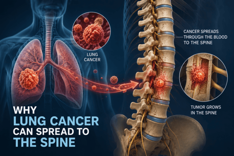
Cancer spreads to the spine via the bloodstream. The vertebral bodies have an extremely rich blood supply through a venous plexus called Batson’s plexus, which connects directly to the venous drainage of the thorax, abdomen, and pelvis. This plexus provides a low-pressure, valve-free pathway for tumour cells to seed the vertebral bones.
Of all the organs where lung cancer metastasises, bone is one of the most common targets, alongside the brain, liver, and adrenal glands. Within bone, the spine, with its extensive and accessible vascular supply, is the single most common site.
Why lung cancer is particularly dangerous in this context:
- Lung cancer is among the most common malignancies worldwide and in the UAE
- It is frequently diagnosed at an advanced stage, when metastatic spread has already occurred
- Vertebral metastases from lung cancer are typically lytic (bone-destroying) rather than blastic (bone-forming), making the affected vertebra structurally weak and prone to collapse
- Collapse or tumour expansion can compress the spinal cord rapidly, as in this case
Other cancers that commonly metastasise to the spine:
- Breast cancer
- Prostate cancer
- Kidney (renal cell) cancer: notably highly vascular, relevant to surgical planning as covered in the earlier article on spinal tumour blood loss
- Thyroid cancer
- Multiple myeloma (haematological malignancy affecting bone marrow)
In any patient with a known history of malignancy who presents with new back pain, spinal metastasis must be actively excluded. Back pain in a cancer patient is a red flag until proven otherwise.
What Metastatic Spinal Cord Compression Means Clinically
Metastatic spinal cord compression (MSCC) is an oncological emergency. It occurs when tumour deposits in the vertebral body expand into the spinal canal, when vertebral collapse from metastatic destruction narrows the canal, or when tumour grows directly within the epidural space.
The spinal cord does not tolerate compression well. When cord function is compromised, the consequences are:
- Motor weakness progressing to paralysis
- Sensory loss below the level of compression
- Bladder and bowel dysfunction
- In complete cord injury: paraplegia or quadriplegia depending on the level
The critical clinical reality is that the neurological outcome of MSCC depends almost entirely on the patient’s neurological status at the time treatment is initiated. A patient who receives decompressive surgery or radiotherapy while still ambulatory has a vastly better outcome than one who has become paraplegic before treatment begins.
Dr. Sherief Elsayed’s immediate response, “I would get an urgent MRI scan of his thoracic spine”, reflects this urgency. Every hour of delay in a patient with rapidly progressive cord compression reduces the likelihood of neurological recovery.
The Neurological Examination: Reading the Signs
The neurological findings in this case deserve individual attention because they are the clinical fingerprint of upper motor neurone cord compression.
Power 2/5 in all lower limb myotomes: Using the Medical Research Council (MRC) grading scale, power of 2 out of 5 means movement is present but cannot overcome gravity. This is severe weakness, below the threshold for useful function. Symmetrical weakness in all lower limb muscle groups indicates a cord-level lesion rather than a nerve root problem, which would produce a specific, asymmetric deficit.
Upgoing plantars (positive Babinski sign): When the sole of the foot is stroked, the normal response is downward flexion of the toes. An upgoing plantar, extension of the big toe, indicates disruption of the descending corticospinal tracts from the brain to the spinal cord. This is an upper motor neurone sign and cannot be produced by a lumbar disc herniation or cauda equina compression.
Pathological clonus: Clonus is rhythmic involuntary muscle contraction in response to sustained stretch. In the ankle, it manifests as repeated bouncing of the foot when the ankle is held in dorsiflexion. Pathological clonus indicates loss of the descending inhibition from the upper motor neurone system, again a cord-level sign.
Hyperreflexia: Exaggerated deep tendon reflexes, as described in the article on Why Spinal Cord Injuries Cause Different Symptoms, reflect the same loss of upper motor neurone inhibition.
Taken together, these four findings constitute an unmistakable upper motor neurone syndrome. They point to the thoracic spinal cord, exactly where the patient has pain. A Spinal Cord Surgeon in Dubai encountering this combination of findings would act with the same urgency.
Treatment of Metastatic Spinal Cord Compression
Treatment depends on the neurological status, the stability of the spine, the tumour type, and the patient’s overall oncological picture.
Immediate medical management:
- High-dose corticosteroids (typically dexamethasone) to reduce cord oedema and slow neurological deterioration while definitive treatment is arranged
- Urgent imaging: whole-spine MRI to identify all metastatic deposits, as multilevel disease is common and affects surgical planning
Surgical decompression:
When the spine is unstable, when there is significant bone involvement, or when the patient is fit enough for surgery, surgical decompression with stabilisation is the most effective way to achieve immediate cord decompression and restore spinal stability. The surgery involves removing the tumour from the epidural space, decompressing the cord, and reconstructing the affected vertebral level with instrumentation.
For the specific challenge of vascular metastases from kidney and thyroid cancer, pre-operative embolisation to reduce blood loss is standard, as covered in the article Why Spinal Tumour Surgery Can Cause Serious Blood Loss.
Radiotherapy:
External beam radiotherapy can treat epidural tumour and reduce cord compression in selected patients, particularly those who are not surgical candidates due to poor fitness. Stereotactic body radiotherapy (SBRT) allows high-dose targeted treatment to vertebral metastases with minimal dose to the adjacent cord.
Outcome:
The single most important determinant of outcome in MSCC is ambulatory status before treatment. Patients who are still walking when treatment begins have a substantially higher chance of remaining ambulatory. Patients who are paraplegic when treatment begins rarely regain independent ambulation. This is the clinical reality that makes the urgency of diagnosis and treatment so critical.
UAE-Specific Context
The UAE has a growing cancer burden, with lung cancer remaining a significant contributor, particularly among older male patients and in the context of historically high smoking rates in the region. Any patient receiving cancer treatment in the UAE who develops new or worsening back pain should be assessed for spinal metastases promptly rather than assuming the pain is musculoskeletal.
The practical point for patients and families: do not accept “muscle strain” as an explanation for new, significant back pain in a patient with known cancer without appropriate investigation. The article When Is Back Pain an Emergency? covers the red flags for urgent spinal assessment in detail, and a cancer history alongside new back pain is one of the most important of them.
Expert Summary
A 45-year-old with lung cancer, bilateral leg weakness, upper motor neurone signs, and thoracic back pain has metastatic spinal cord compression until proven otherwise. The wrong investigation. A lumbar MRI that does not image the thoracic spine, provides false reassurance when the clinical signs are pointing elsewhere. The right investigation, an urgent thoracic MRI, confirms the diagnosis and opens the door to treatment before neurological damage becomes permanent.
Dr. Sherief Elsayed’s clinical reasoning illustrates something that applies broadly in spine medicine: the scan must be directed by the clinical signs, not chosen by default. For any patient in Dubai with a history of malignancy and new spinal symptoms, consulting a Spine Tumour Surgeon in Dubai without delay is the most important step toward preserving neurological function.
Table of Contents
Recent Articles

Why Eight People Were Needed for One Spine Surgery in Dubai
Why Eight People Were Needed for One Spine Surgery in Dubai Does every spinal surgery require exactly eight team members? No, as Dr. Sherief Elsayed explains directly, the specific team

Three Spine Surgery Myths a Dubai Surgeon Wants You to Forget
Three Spine Surgery Myths a Dubai Surgeon Wants You to Forget Is it really safe to walk on the same day as spine surgery? Yes, for the majority of modern

Why Osteoporosis Is Called the Silent Disease in the UAE
Why Osteoporosis Is Called the Silent Disease in the UAE If osteoporosis doesn’t cause pain, how would I know if I needed a DEXA scan? Given the entirely silent nature