
Why Most Modern Pacemakers Are Now MRI Compatible in Dubai
How do I find out if my specific pacemaker is MRI compatible?
Your cardiologist or the pacemaker manufacturer can confirm the specific compatibility status of both your device generator and your leads. Many patients are also given a device identification card at the time of implantation that includes this information.
Is there any risk during the MRI scan itself, even with an MRI-conditional pacemaker?
When appropriate protocols are followed, including confirming device and lead compatibility and correctly programming the MRI-safe mode beforehand, the risk is very low. The scanning facility and cardiology team follow specific established safety protocols for exactly this scenario.
Can the pacemaker programming settings be affected permanently by an MRI scan?
No, when the correct process is followed, the temporary MRI-safe mode is specifically designed to be reversible, and the device is reprogrammed back to its normal settings immediately after the scan is completed.
What should I do if I need an MRI urgently and I am not sure about my pacemaker's compatibility?
Inform the treating medical team immediately about your pacemaker, and they will coordinate with cardiology to determine compatibility and, if needed, discuss whether an alternative imaging modality such as CT might be appropriate in the interim.
Are all types of implanted cardiac devices covered by this same MRI compatibility advance, not just pacemakers?
Implantable cardioverter-defibrillators (ICDs) and other cardiac devices have similarly seen the development of MRI-conditional models, though the specific compatibility status needs to be confirmed individually for each device type and model, following the same principle of checking both the generator and leads.
How long does the process of switching the pacemaker into MRI mode and back take?
This is typically a quick process performed by the cardiology team or pacemaker technician using a wireless programming device, usually taking only a few minutes before and after the scan itself.
For many years, patients with pacemakers were told a simple, absolute rule: no MRI scans, ever. This was genuinely sound advice at the time, reflecting real risks with older pacemaker technology. That rule has changed significantly, and the persistence of outdated advice means that patients today are sometimes denied access to the best available imaging for their condition based on information that no longer reflects current pacemaker technology.
Dr. Sherief Elsayed, Consultant Spine Surgeon in Dubai, addresses this directly and explains what has actually changed.
The Outdated Assumption
Dr. Sherief Elsayed identifies the common misconception directly: “A lot of patients mistakenly think that if they have a pacemaker, they can’t have an MRI scan.”
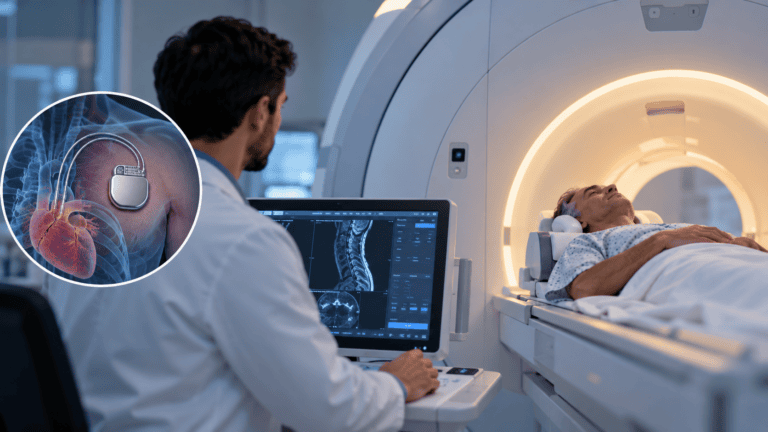
This assumption was, for decades, entirely correct. Older generation pacemakers contained components that could malfunction in the powerful magnetic field of an MRI scanner. The magnetic field could induce electrical currents in the pacemaker leads, potentially causing unintended cardiac stimulation, heating at the lead tips that could damage surrounding heart tissue, or malfunction of the pacemaker’s programming, any of which could be genuinely dangerous for a patient dependent on the device for normal heart rhythm.
Given these real historical risks, the blanket “no MRI” rule was appropriate advice for a long time, and it has understandably persisted in general public awareness even as the underlying technology has moved on considerably.
What Has Actually Changed
Dr. Sherief Elsayed states the current reality plainly: “Whereas actually, most modern pacemakers are MRI compatible.”
Pacemaker manufacturers have specifically engineered newer generation devices to address the risks that made older pacemakers incompatible with MRI. These modern devices, often marketed as “MRI conditional” pacemakers, incorporate design features including modified lead materials and configurations that reduce the risk of induced heating, circuitry designed to withstand the electromagnetic environment of an MRI scanner without malfunctioning, and a specific “MRI mode” that can be activated before a scan to further reduce any residual risk.
This is a genuinely significant advance, given how common both pacemaker implantation and MRI scanning are, particularly in an ageing population where the two clinical needs frequently overlap. A patient with a pacemaker who develops a spinal condition requiring MRI assessment, a scenario directly relevant to the population Dr. Sherief Elsayed treats, previously faced a genuine diagnostic dilemma. Modern pacemaker technology has substantially resolved this for the majority of patients.
The Two Things That Must Be Confirmed
Dr. Sherief Elsayed is specific about what needs to be verified before proceeding: “You need to make sure that the device is compatible and the leads going to your heart are compatible.”
This distinction matters because MRI compatibility is not simply a property of the pacemaker generator (the small device implanted under the skin, usually near the collarbone) in isolation. The leads, the thin insulated wires that run from the generator to the heart itself, are an equally important component of the system, and older leads connected to a newer, MRI-compatible generator do not automatically make the overall system safe for MRI scanning.
Device compatibility: The pacemaker generator itself must be a model specifically designed and certified as MRI conditional by its manufacturer. This information is recorded in the patient’s device documentation and can be confirmed through the cardiology team or pacemaker manufacturer’s records.
Lead compatibility: The specific leads connecting the generator to the heart must also be certified as MRI conditional. In some patients, particularly those who have had their pacemaker generator replaced or upgraded over time while retaining their original leads, there can be a mismatch, a fully MRI-compatible modern generator connected to older, non-MRI-compatible leads, meaning the overall system does not meet the safety criteria for MRI scanning despite the generator itself being a compatible model.
The Practical Process: What Happens Before and After the Scan
Dr. Sherief Elsayed outlines the specific clinical pathway: “What happens is you see your cardiologist or the pacemaker technician who will place it into MRI mode and go have your MRI scan. And then as soon as the scan’s finished, it’s placed back into its regular mode.”
Before the scan:
A cardiologist or specialist pacemaker technician uses a programming device to communicate wirelessly with the implanted pacemaker and switch it into a specific MRI-safe mode. This mode typically adjusts the pacemaker’s settings, for instance, switching to an asynchronous pacing mode that is less susceptible to any electromagnetic interference from the scanner, and may disable certain sensing functions that could be temporarily affected by the MRI environment.
During the scan:
With the device correctly programmed into MRI mode, and following the specific scanning protocols and parameter limits established for that particular device model, the patient can safely undergo the MRI scan.
After the scan:
Once scanning is complete, the same cardiologist or technician reprograms the device back into its normal, regular operating mode, restoring the full range of pacing and sensing functions that were temporarily adjusted for the MRI-safe mode.
This process requires coordination between the radiology team performing the scan and the cardiology team managing the pacemaker, which is why patients with pacemakers requiring MRI need this specifically arranged in advance rather than simply booking a routine MRI appointment.
Why Dr. Sherief Elsayed’s Caveat Matters
Despite the genuinely positive shift in pacemaker MRI compatibility, Dr. Sherief Elsayed is careful not to overstate the universality of this advance: “Now that’s not suitable for everybody, but ask your cardiologist.”
This caution is clinically important. Not every pacemaker in current use is an MRI-conditional model. Patients with older devices that have not yet required replacement, patients with non-MRI-compatible leads even alongside a newer generator, and patients with certain other implanted cardiac devices that fall outside standard MRI-conditional certification, may still face genuine restrictions on MRI scanning.
Additionally, even for patients with fully MRI-conditional systems, specific scanning parameters, the strength of the magnetic field, the specific body region being scanned, and other technical factors, may still need to be confirmed as falling within the certified safe parameters for that particular device model.
Why This Matters Specifically in Spine Care
MRI is the primary imaging modality for the vast majority of spinal conditions. A Spine Surgeon in Dubai will coordinate directly with the cardiology team to determine MRI eligibility for pacemaker patients. discussed throughout this blog, from disc herniation and spinal stenosis to spinal cord compression and tumour assessment. A patient with a pacemaker who develops significant back or neck pain, or neurological symptoms suggesting a spinal cause, needs the same quality of diagnostic imaging as any other patient to reach an accurate diagnosis and appropriate treatment plan.
Before the widespread availability of MRI-conditional pacemakers, patients in this situation often had to rely on CT scanning as an alternative, which, while valuable for assessing bony anatomy, provides significantly less detailed information about the soft tissue structures, including discs, nerve roots, and the spinal cord itself, that are frequently central to diagnosing and planning treatment for spinal conditions. A Spine Doctor in Dubai recognises how significantly this affects the quality and completeness of diagnostic information available.
A Spine Surgeon in UAE assessing a patient with a pacemaker and a suspected spinal condition will specifically coordinate with the patient’s cardiology team to determine MRI eligibility, recognising how significantly this affects the quality and completeness of the diagnostic information available.
UAE-Specific Context
Major hospitals and specialist cardiac and imaging centres across Dubai and the UAE are equipped with the necessary MRI scanning technology. A Consultant Spine Surgeon in Dubai works within this infrastructure to ensure pacemaker patients receive appropriate imaging. and the UAE are equipped with the necessary MRI scanning technology and cardiology support to safely manage MRI-conditional pacemaker patients through this coordinated process. Given the UAE’s significant expatriate population, some of whom may have had their pacemaker implanted in a different country, ensuring that complete and accurate device and lead compatibility documentation is available, ideally through direct communication between the treating cardiology team and the original implanting centre where needed, is an important practical step in facilitating safe MRI access.
Expert Summary
Most modern pacemakers are now MRI compatible, representing a genuine and clinically significant advance from the older, absolute restriction that many patients and even some healthcare providers still assume applies universally. Both the pacemaker generator and its connecting leads must be confirmed as MRI conditional, and the scanning process requires the device to be switched into a specific MRI-safe mode beforehand and returned to normal mode immediately afterward. This is not suitable for every pacemaker patient, making a direct conversation with the treating cardiologist an essential step before assuming either that MRI is impossible or that it is automatically safe.
Table of Contents
Recent Articles

Why Eight People Were Needed for One Spine Surgery in Dubai
Why Eight People Were Needed for One Spine Surgery in Dubai Does every spinal surgery require exactly eight team members? No, as Dr. Sherief Elsayed explains directly, the specific team
Three Spine Surgery Myths a Dubai Surgeon Wants You to Forget
Three Spine Surgery Myths a Dubai Surgeon Wants You to Forget Is it really safe to walk on the same day as spine surgery? Yes, for the majority of modern
Why Osteoporosis Is Called the Silent Disease in the UAE
Why Osteoporosis Is Called the Silent Disease in the UAE If osteoporosis doesn’t cause pain, how would I know if I needed a DEXA scan? Given the entirely silent nature