
Why Bone Cement Is Injected Into the Spine and What the Risks Are
Is bone cement permanent?
Yes. PMMA bone cement is not absorbed or metabolised by the body. Once hardened, it remains in place permanently. It does not integrate biologically with surrounding bone in the way that true bone graft does, but it provides durable mechanical support.
Is the cement injection procedure painful?
Vertebroplasty and kyphoplasty are typically performed under local anaesthesia with sedation for pain control. Most patients experience some discomfort during the procedure but not severe pain. Post-procedure soreness at the injection site usually resolves within a few days. Significant pain relief from the stabilised fracture is often reported within hours to days of the procedure
How quickly can I return to normal activity after vertebroplasty?
Most patients can mobilise and be discharged within hours of the procedure. Return to light activity is typically within a few days, with full activity guided by the surgeon’s advice and the patient’s overall condition. The structural stabilisation is immediate.
Will the cement show on X-ray or MRI?
PMMA cement is very clearly visible on X-ray and CT because it is radiopaque. On MRI, it is also identifiable as a low-signal (dark) area within the vertebra, and does not produce the same degree of artefact as metallic implants. This allows the radiologist to assess the distribution of cement and the surrounding bone on post-operative imaging.
What happens if cement leaks outside the vertebra?
Cement leakage is the most common complication and is typically detected on intraoperative imaging. Small amounts of leakage into the disc space or paraspinal tissue are usually clinically inconsequential. Leakage into the spinal canal or foramina, compressing neural structures, is a more serious complication that may require surgical intervention. This risk is minimised by fluoroscopic guidance during injection and the use of appropriate cement viscosity.
Is kyphoplasty better than vertebroplasty?
The two procedures share the same principle of cement injection but differ in technique and clinical goals. Kyphoplasty’s balloon inflation step offers the potential for partial vertebral height restoration and creates a cavity that allows cement injection at lower pressure, potentially reducing extravasation risk. For acute fractures where height restoration is a priority, kyphoplasty may be preferable. For established fractures where height restoration is unlikely, the additional step of balloon inflation may add less benefit. The choice is made on an individual basis.
Bone cement is one of the more counterintuitive interventions in spine surgery. The idea of injecting a liquid polymer directly into a vertebra, where it then hardens in place, sounds dramatic and perhaps alarming to patients who are unfamiliar with the technique. In practice, it is a precise and clinically important tool for specific situations, particularly where structural bone loss has compromised the integrity of a vertebra and conventional implant fixation alone is insufficient.
Dr. Sherief Elsayed, Consultant Spine Surgeon in Dubai, describes using bone cement directly from an operative case involving a vertebral tumour at T11, explaining both the volume used, the rationale, and the specific risk that every patient receiving this treatment should understand.
The Case: Tumour Reconstruction at T11
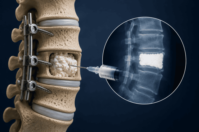
Dr. Sherief Elsayed narrates the intraoperative findings: “I can see the patient’s spine from the side. Here is where the tumour was. I filled that area with cement and then I’ve supplemented the whole thing with screws so that there’s no collapse. This instrument shows that I’ve decompressed below and above the level of the T11 vertebra. Total volume of just under 18 mLs of cement. Quite a large volume, but he’s a very big gentleman and his vertebra needed that much.”
Several important clinical points are embedded in this description.
Why the tumour cavity required cement:
When a tumour is removed from a vertebral body, it leaves behind a structural defect. The vertebra, already weakened by the tumour’s presence, now has a cavity where bone material previously existed. Without filling this cavity, the vertebra cannot adequately support the compressive loads of the spinal column. Collapse and progressive deformity would follow, with potential consequences for neural structures.
Bone cement, or polymethylmethacrylate (PMMA), fills the cavity immediately and hardens within minutes, restoring the structural integrity of the vertebral body. This is supplemented, as in this case, with pedicle screws and rods that provide additional stability to the reconstructed segment.
Why 18 millilitres was a large but appropriate volume:
The amount of cement used is proportional to the size of the vertebral defect and the patient’s anatomy. A larger vertebra in a larger patient requires a proportionally larger volume. A Spinal Reconstruction Surgeon in Dubai determines the appropriate cement volume based on individual anatomy and the extent of the structural defect. to achieve adequate fill. The volume must be sufficient to fill the structural defect, but not so excessive that it risks extravasation, which refers to cement leaking beyond the intended boundaries into surrounding structures.
What Is Bone Cement and How Does It Work?
Polymethylmethacrylate bone cement has been used in orthopaedic and spinal surgery for decades, originally developed for fixation of joint replacement implants and later adapted for vertebral augmentation procedures.
Its properties:
PMMA is supplied as a powder and liquid that are mixed at the point of use. The mixture is injectable for a defined working time, during which it can be directed into the target location. Once the working time expires, the cement undergoes an exothermic polymerisation reaction and hardens into a rigid, stable material that can immediately bear compressive load.
Its clinical advantages:
It provides immediate mechanical stability at the treated level, without waiting for biological healing. It fills irregular cavities that conventional implants cannot address. It distributes compressive load across the vertebral end plate, reducing the risk of further collapse. In the context of malignant disease, the heat generated during hardening may also have a local anti-tumour effect.
Where Else Is Bone Cement Used in the Spine?
Beyond tumour reconstruction, bone cement is used in two common vertebral augmentation procedures.
Vertebroplasty: Cement is injected directly into an osteoporotic vertebral compression fracture under imaging guidance. The procedure stabilises the fractured vertebra, reduces pain, and prevents further collapse. It is typically performed as a minimally invasive procedure under local anaesthesia and sedation.
Kyphoplasty: A balloon is first introduced into the fractured vertebra and inflated to partially restore vertebral height and create a cavity. Cement is then injected into this cavity at lower pressure, reducing the risk of cement extravasation compared to vertebroplasty. The restored cavity also provides an opportunity for partial height restoration before the cement hardens.
Both procedures address the painful, structurally compromised vertebra without the need for open surgery or general anaesthesia in most cases. A Spine Fusion Doctor in Dubai can advise whether vertebroplasty, kyphoplasty, or a more extensive reconstruction is appropriate for a given fracture pattern. without the need for open surgery or general anaesthesia in most cases.
The Risk That Must Be Understood: Marrow Embolisation
Dr. Sherief Elsayed is direct about the specific risk of bone cement injection: “The problem when you inject bone cement into a vertebra is marrow embolisation. The marrow gets spread out and can cause problems in the lungs, for example.”
This risk deserves careful explanation, because understanding it is part of genuinely informed consent for any procedure involving bone cement in the spine.
What marrow embolisation is:
The vertebral body contains bone marrow within a network of vascular channels. When cement is injected under pressure into this space, it displaces the marrow contents. Marrow fat, cells, and other material can be forced into the vertebral venous plexus, the same rich network of veins through which spinal tumours characteristically seed, as described in the lung cancer article in this series. From the venous system, this material can travel to the right side of the heart and into the pulmonary circulation.
If sufficient embolised material reaches the lung vasculature, it can cause fat embolism syndrome or pulmonary embolism, ranging in severity from clinically silent (no symptoms) to life-threatening respiratory compromise.
How the risk is managed:
Several strategies reduce the risk of clinically significant marrow embolisation during bone cement procedures.
Cement is injected slowly and under fluoroscopic or CT guidance so that the radiologist or surgeon can watch in real time as the cement fills the cavity and immediately stop if extravasation or venous runoff is detected. Cement viscosity is matched to the procedure: a thicker, more viscous cement is harder to inject but less prone to flowing into vascular channels. The volume injected is kept to the minimum necessary for structural stability. Intraoperative cardiac monitoring detects early haemodynamic changes that may indicate significant embolisation, allowing the team to pause.
The absolute incidence of clinically significant pulmonary embolism from bone cement procedures is low when performed by experienced practitioners with appropriate monitoring. However, it is a genuine risk that should be disclosed to every patient receiving this treatment, and the case described by Dr. Sherief Elsayed, with a relatively large volume in a complex tumour reconstruction case, represents exactly the situation where this risk is most relevant.
Who Are the Best Candidates for Bone Cement Procedures?
Patient selection is the most important determinant of outcome and safety for cement-based spinal procedures.
Well-suited candidates include:
Patients with osteoporotic vertebral compression fractures causing significant pain that has not responded to conservative management. The fracture should be acute or subacute (within the past few months) and must still have viable potential for height restoration in the case of kyphoplasty. Patients with pathological fractures from spinal metastases in whom structural reconstruction is needed alongside tumour treatment.
Less suitable or requiring careful assessment:
Patients with fractures involving the posterior vertebral wall, where cement injection carries a higher risk of posterior extravasation into the spinal canal and cord compression. Patients with significant coagulopathy that increases bleeding risk. Patients with active infection at the target level. Cases where the fracture has already collapsed to a degree that makes meaningful height restoration unlikely.
A Spine Tumour Surgeon in Dubai assessing a patient with a metastatic vertebral lesion for cement augmentation will weigh the structural benefit of the procedure against the risk of marrow embolisation and other complications, and will obtain imaging guidance support from interventional radiology to ensure the procedure is performed with maximum precision and safety.
UAE-Specific Context
Vertebroplasty and kyphoplasty for osteoporotic fractures are available at major spinal centres across Dubai and the UAE. With the UAE’s high rates of vitamin D deficiency, sedentary lifestyle prevalence, and an ageing population, the burden of osteoporotic vertebral fractures in the region is significant. Awareness of these procedures as options for painful, compression-fractured vertebrae is important for both patients and their primary care physicians, many of whom may not be familiar with interventional spinal options beyond conservative management.
A Consultant Spine Surgeon in Dubai who performs these procedures routinely can explain precisely what they involve, what the realistic benefits and risks are for an individual patient’s anatomy and fracture pattern, and what monitoring will be in place during the procedure to manage the risk of cement complications.
Expert Summary
Bone cement fills a specific and important clinical gap in spinal surgery: the immediate restoration of structural integrity to vertebrae that have been compromised by tumour, osteoporotic fracture, or other structural pathology, without waiting for biological healing. It is effective, technically established, and in experienced hands, very safe. The risk of marrow embolisation, where cement injection displaces vertebral marrow into the pulmonary circulation, is real and requires careful technique, appropriate imaging guidance, and intraoperative monitoring. Disclosed honestly and managed carefully, it is a risk that is clinically justified by the structural benefit in well-selected patients.
Table of Contents
Recent Articles

Why Eight People Were Needed for One Spine Surgery in Dubai
Why Eight People Were Needed for One Spine Surgery in Dubai Does every spinal surgery require exactly eight team members? No, as Dr. Sherief Elsayed explains directly, the specific team
Three Spine Surgery Myths a Dubai Surgeon Wants You to Forget
Three Spine Surgery Myths a Dubai Surgeon Wants You to Forget Is it really safe to walk on the same day as spine surgery? Yes, for the majority of modern
Why Osteoporosis Is Called the Silent Disease in the UAE
Why Osteoporosis Is Called the Silent Disease in the UAE If osteoporosis doesn’t cause pain, how would I know if I needed a DEXA scan? Given the entirely silent nature