What Is Spinal Derotation and Why Is It Key to Scoliosis Surgery in the UAE
Will I be able to bend normally after scoliosis surgery?
This depends on the levels fused. Patients with thoracic-only fusions retain good lumbar mobility. Patients with long fusions extending into the lower lumbar spine will have reduced trunk rotation and lateral bending but typically adapt well and maintain good functional movement for activities of daily living.
How long do the implants stay in?
In the majority of patients, the pedicle screws and rods remain in place permanently. They are not routinely removed once the fusion has consolidated. Removal is considered only if the implants cause specific problems such as pain from prominent metalwork or infection.
Is the surgery painful?
Post-operative pain is managed with a multimodal analgesia protocol including regular oral pain medication, anti-inflammatory agents, and nerve pain medication where appropriate. Most patients describe the pain as manageable rather than severe, and it improves progressively over the first two to four weeks.
What are the risks of scoliosis surgery?
The main risks include infection, bleeding, neurological injury (rare but serious), implant failure, failure to achieve fusion (pseudoarthrosis), and adjacent segment degeneration over the long term. Modern neuromonitoring and surgical technique have substantially reduced the risk of neurological complications. Your surgeon will discuss the specific risk profile for your case at the consent consultation.
Can scoliosis come back after surgery?
Once the instrumented levels are fully fused, the correction at those levels is permanent. The spine cannot re-curve through the fused segment. However, adjacent unfused levels can develop their own changes over time, which is why the choice of fusion levels is made carefully to include all curve components that require stabilisation.
Is there an age limit for scoliosis surgery?
There is no strict upper age limit. Surgery for adult degenerative scoliosis is performed in appropriately selected patients of advanced age. The key factors are the patient’s overall health, bone quality, and the balance between the expected benefit of surgery and the risks associated with the procedure in that individual. Thorough pre-operative assessment determines surgical suitability on a case-by-case basis.
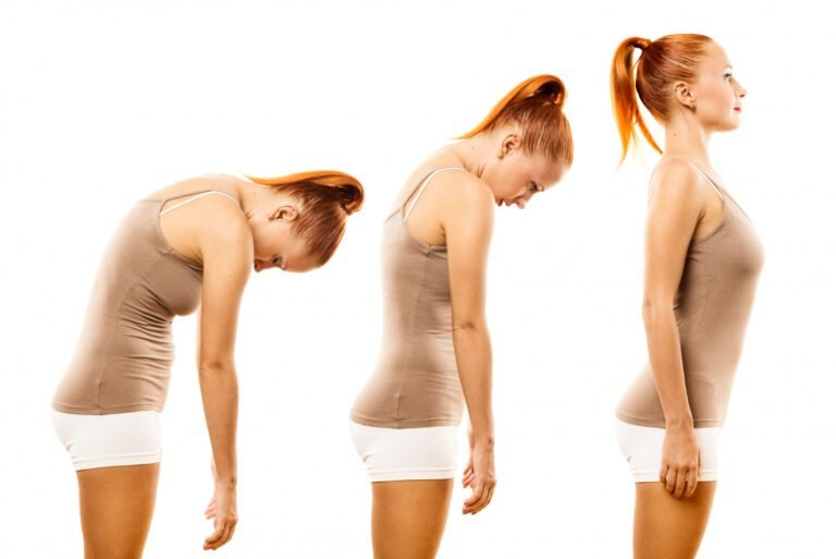
Scoliosis is not simply a curve. That is the most common misconception about the condition, and it matters clinically because surgery that addresses only the lateral bend without correcting the rotational deformity produces an incomplete result. The vertebrae in a scoliotic spine do not just tilt sideways, they also rotate, creating the characteristic rib hump that is often the most visible cosmetic feature of the condition. Correcting scoliosis properly means correcting both the curve and the rotation simultaneously, and the technique used to achieve that is one of the most technically demanding manoeuvres in spinal surgery.
Dr. Sherief Elsayed, Consultant Spine Surgeon in Dubai, describes the surgical sequence for scoliosis correction in precise detail, including the derotation manoeuvre that is central to achieving the best possible outcome.
Why Is Scoliosis a Three-Dimensional Deformity?
Understanding why derotation matters requires a brief explanation of scoliosis mechanics.
In a healthy spine viewed from the front, the vertebrae are stacked symmetrically. In adolescent idiopathic scoliosis, the most common form, the vertebrae gradually rotate as the curve develops. Because the ribs attach to the thoracic vertebrae, vertebral rotation pulls the ribs on the convex side of the curve posteriorly, creating the rib prominence or rib hump that is often the first sign a parent notices when a child bends forward. On the concave side, the anterior chest wall may appear flattened or hollow.
This rotational component cannot be corrected by simply pulling the spine straighter in the coronal (front-to-back) plane. A system that straightens the curve without derotating the vertebrae will improve the X-ray appearance but leave the rib hump largely unchanged, which is often the patient’s primary cosmetic concern.
Modern scoliosis instrumentation and surgical techniques address all three planes of the deformity, correcting the lateral curve, the sagittal profile (the forward and backward curvature), and the axial rotation simultaneously.
The Surgical Sequence: What Actually Happens in the Operating Theatre
Dr. Sherief Elsayed describes the operative steps with the clarity of someone who has performed this procedure many times: “First, we put all the screws into the levels that we want to. Next, we contour a rod to the shape that we want the spine to end up as at the end. We place that down onto the screws and you’ll see that it doesn’t quite fit right. So then we add our reduction towers and that helps to force the rod down onto the screws before we do our derotation manoeuvre, which straightens out the spine. We then fix the rod to the screws from top to bottom before we apply the next rod followed by a bone fusion and then closure.”
This description outlines a sequence that reflects the evolution of scoliosis surgery over decades of technical refinement.
Step 1: Pedicle Screw Placement
Pedicle screws are placed into each vertebra at the levels to be included in the fusion. The pedicle is the bony bridge connecting the posterior elements of the vertebra to the vertebral body, and it provides the strongest possible purchase for instrumentation. Screws placed through both pedicles at each level create bilateral anchor points through which corrective forces can be applied.
The accuracy of screw placement is critical. Misplaced pedicle screws can breach the spinal canal or exit the pedicle toward adjacent neural or vascular structures. Modern scoliosis surgery uses intraoperative imaging, navigation, and neuromonitoring to ensure screw placement is precise and safe throughout.
Step 2: Rod Contouring
Before the rod is inserted, it is bent to match the three-dimensional shape that the surgeon intends the spine to adopt at the end of the procedure. This is not a flat bend but a complex contoured shape that accounts for the desired coronal correction, the sagittal profile, and the derotation that will follow.
The rod essentially serves as a template for the corrected spine. When it is fully seated and locked, the spine will conform to the shape of the rod rather than the rod conforming to the shape of the spine.
Step 3: Rod Seating and Reduction Towers
As Dr. Sherief Elsayed notes, the contoured rod does not drop easily into the screw heads when first placed because the screws are currently positioned in the deformed configuration. Reduction towers are specialised instruments that attach to individual screw heads and allow the surgeon to lever the rod down into each screw progressively, translating the vertebra toward the rod and beginning the correction.
This process, systematically working along the construct from one end to the other, gradually brings the deformed spine toward the corrected position defined by the rod.
Step 4: The Derotation Manoeuvre
This is the most technically significant step and the one that addresses the rotational component of the deformity. Once the rod is seated, derotation instruments are applied to the rod or to specialised towers attached to key screw heads. By rotating the rod in a controlled manner, the vertebrae attached to the screws are carried with it, derotating the spine in the axial plane.
The rib hump, which is the external visible manifestation of the vertebral rotation, reduces as the derotation is performed. The surgical team monitors the correction radiographically throughout, and neuromonitoring of the spinal cord and nerve roots runs continuously to ensure that the corrective manoeuvres do not compromise neurological function.
A Scoliosis Specialist in Dubai (Scoliosis Treatment in Dubai – Adult & Paediatric) who performs this procedure regularly will have the technical experience to achieve the maximum safe correction while protecting neurological function throughout.
Step 5: Final Rod Fixation and Second Rod Application
Once the derotation is complete and the correction is confirmed on intraoperative imaging, the rod is locked to each screw head sequentially from top to bottom. A second rod is then applied on the contralateral side, which provides rotational stability to the construct and prevents the correction from unwinding.
The dual rod construct, one on each side of the spine, creates a mechanically stable fixation system that holds the correction while the bone graft consolidates into a solid fusion.
Step 6: Bone Graft and Fusion
Decortication of the posterior elements creates a bleeding bone surface that accepts the graft material. Bone graft, either harvested locally from the surgical site, supplemented with allograft (donor bone), or combined with bone graft substitutes, is packed along the posterior elements across all the instrumented levels. Over the following months, this graft consolidates into a solid bridge of bone that permanently maintains the correction. A Spinal Fusion Doctor in Dubai (Spinal Conditions – Diagnosis & Treatment in Dubai) will monitor this healing process with regular reviews and imaging throughout your recovery.
Step 7: Closure
The wound is closed in anatomical layers, with meticulous attention to haemostasis. A drain is typically placed to prevent haematoma formation in the post-operative period.
Why Neuromonitoring Is Non-Negotiable in Scoliosis Surgery
Scoliosis correction places the spinal cord and nerve roots under mechanical stress. The derotation manoeuvre in particular, while essential to the best cosmetic and functional outcome, involves forces applied to a spine in which the cord has adapted to a deformed position over years.
Intraoperative neuromonitoring, using a combination of somatosensory evoked potentials (SSEPs) and motor evoked potentials (MEPs), tracks spinal cord function continuously throughout the procedure. Any change in signal quality alerts the surgical team immediately, allowing them to pause, reassess, and modify their approach before a neurological injury becomes established.
At leading spinal surgical centres, neuromonitoring has become the standard of care for scoliosis correction. Its use does not eliminate neurological risk entirely, but it provides a real-time safety net that has significantly reduced the incidence of post-operative neurological deficits in scoliosis surgery.
What Results Can Patients Expect From Scoliosis Correction?
Modern instrumented scoliosis correction achieves substantial curve reduction in the majority of patients. Curves that entered surgery at 50 to 60 degrees are routinely corrected to under 20 degrees, representing a 60 to 70 percent correction. The cosmetic improvement, with reduction of the rib hump and restoration of trunk balance, is often the most meaningful outcome for patients who have been self-conscious about their appearance.
Realistic expectations include:
- Significant reduction in the curve angle, typically 60 to 70 percent correction
- Meaningful reduction in the rib hump, particularly when derotation is achieved
- Restoration of trunk balance and horizontal shoulder alignment
- Prevention of further progression
- Improvement in back pain where it was a significant pre-operative symptom, though pain relief is less predictable than curve correction
The fusion levels chosen determine what movements the spine retains after surgery. Selective thoracic fusion preserves lumbar mobility in patients where the lumbar curve is flexible and non-structural. Long fusions extending into the lumbar spine reduce rotational and lateral mobility but are necessary in patients with structural lumbar curves.
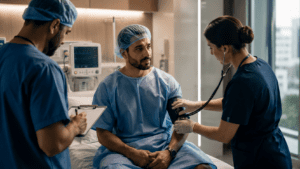
Recovery After Scoliosis Correction Surgery
Most patients are mobilised and walking within 24 to 48 hours of surgery. The procedure itself typically takes three to six hours depending on the number of levels fused and the complexity of the deformity. Hospital stay is generally four to seven days.
Typical recovery milestones:
- Return to school or light activity: four to six weeks. To understand what your specific correction will involve, a Scoliosis Correction Surgeon in Dubai (Scoliosis Treatment in Dubai – Adult & Paediatric) can walk you through the full surgical and recovery process.
- Return to non-contact sport: three to six months
- Return to contact sport or heavy physical activity: twelve months
- Full fusion consolidation: twelve to eighteen months
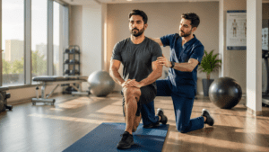
Post-operative physiotherapy focuses on core strengthening, posture, and graduated return to activity rather than attempting to alter the correction, which is fixed by the instrumentation.
Expert Summary
Scoliosis surgery is not simply bending a rod and attaching it to the spine. It is a technically precise, sequenced procedure that addresses a three-dimensional deformity through careful screw placement, rod contouring, reduction, derotation, and fusion. The derotation manoeuvre is the step that separates a good cosmetic result from a merely adequate radiographic one, and it requires the experience and technical precision of a surgeon who performs this procedure regularly. For patients in the UAE facing scoliosis correction, understanding what the surgery involves, and why each step matters, makes the entire experience less intimidating and more meaningful. To discuss your specific curve and what correction is achievable, consulting a Spine Surgeon in UAE (Spinal Conditions – Diagnosis & Treatment in Dubai) with dedicated scoliosis experience is the right starting point.
Table of Contents
Recent Articles
Why 15% of Disc Surgery Patients Get a Recurrence Within a Year in Dubai
Why 15% of Disc Surgery Patients Get a Recurrence Within a Year in Dubai Is recurrent disc prolapse more serious than the original prolapse? Not necessarily. The recurrent herniation may
Why Low Blood Pressure Can Delay Your Surgery in the UAE
Why Low Blood Pressure Can Delay Your Surgery in the UAE Can anxiety cause low blood pressure before surgery? Yes. Anxiety activates the sympathetic nervous system, which typically raises blood

When Do You Actually Need an Antibiotic? A UAE Doctor Explains
When Do You Actually Need an Antibiotic? A UAE Doctor Explains My doctor prescribed an antibiotic for my cold. Was that wrong? Almost certainly yes. The common cold is a