
What Happens When a Spine Cage Is Placed in the Wrong Position?
How is cage malpositioning diagnosed?
Post-operative imaging is essential. Plain X-rays can identify obvious cage malpositioning. CT scanning provides the most detailed assessment of cage position relative to the posterior vertebral wall, spinal canal, and foramina, and is the standard investigation when malpositioning is suspected. MRI may show neural compression and is useful for assessing the degree of neural involvement.
Can cage malpositioning be treated without surgery?
If malpositioning is not causing neural compression or symptoms, conservative monitoring may be appropriate. However, when malpositioning is causing neurological symptoms, revision surgery is generally the appropriate treatment because removing the structural cause of compression is the only way to reliably resolve neurologically mediated symptoms.
How long after the original surgery can revision be performed?
Revision is most technically straightforward in the early post-operative period before significant adhesion formation. However, it can be performed at any point when symptoms indicate the need, including months after the original procedure. The technical challenge increases with time from the original surgery.
What are the risks of revision ALIF surgery?
Revision anterior lumbar surgery carries higher risks than primary surgery, primarily related to adhesion and fibrosis around the major blood vessels mobilised during the original procedure. Vascular injury, though uncommon in experienced hands, is a more significant risk in revision cases, which is why surgical teams with available vascular support are preferred for these procedures.
Is cage malpositioning a common complication?
Significant malpositioning causing neural compression is an uncommon complication. Minor deviations from ideal cage positioning that do not cause symptoms are more common and typically do not require revision. The incidence of clinically significant malpositioning is reduced by intraoperative three-dimensional imaging verification of cage position.
What questions should I ask before an ALIF procedure?
Ask whether intraoperative three-dimensional imaging (O-arm or equivalent) will be used to verify cage position before wound closure. Ask whether posterior supplemental fixation is planned and in what sequence. Ask about the surgeon’s experience with both primary ALIF and revision procedures, as the latter experience indicates familiarity with the full spectrum of outcomes.
Spinal surgery is a precise discipline. The implants used in fusion procedures, specifically the cages placed within disc spaces to restore height and provide a scaffold for fusion, must be positioned within narrow anatomical boundaries to achieve their intended purpose safely. When a cage is placed incorrectly, the consequences range from suboptimal fusion to serious neurological injury. Understanding what correct cage placement looks like, why it sometimes goes wrong, and what can be done about it, is part of what informed patients and referring clinicians should know.
Dr. Sherief Elsayed, Consultant Spine Surgeon in Dubai, describes a case that illustrates what a mispositioned cage looks like on imaging and what the appropriate clinical response is.
The Case: An ALIF Cage in the Wrong Position
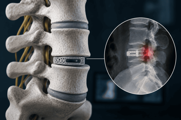
Dr. Sherief Elsayed presents the imaging: “This is a post-operative X-ray with an ALIF cage in the wrong position. ALIF is a procedure where we come in from the front, and it’s an anterior lumbar interbody fusion. You can see this is a cage, but it’s too posterior. It’s gone too far back. It should be sitting nice and centrally. This cage has gone into the posterior wall of the vertebra and is now causing nerve compression and pain.”
This description identifies several important clinical elements simultaneously.
What “too posterior” means:
An ALIF cage is placed through an anterior (front of the abdomen) approach into the disc space between two adjacent vertebrae. The correct position is anterior to central within the disc space, away from the posterior vertebral wall and the spinal canal and nerve roots that lie directly behind it. A Spine Fusion Doctor in Dubai performing an ALIF procedure verifies this position carefully before completing the surgery.
When a cage migrates posteriorly, or is initially placed too far back, its trailing edge impacts the posterior vertebral wall and may protrude into or through the posterior cortex, directly compressing the nerve roots in the lateral recess or the thecal sac centrally.
The symptom consequence:
A cage that has compressed neural structures will produce either radicular pain (if a specific nerve root is compressed, producing leg pain in a specific distribution) or bilateral lower limb symptoms or bladder disturbance (if central compression of the cauda equina is present). The patient’s symptoms directly reflect the degree and pattern of neural compression from the malpositioned cage.
Why Cage Malpositioning Happens
Several mechanisms can contribute to cage malpositioning during or after ALIF or other interbody fusion procedures.
Technical factors during insertion:
The anterior approach to the lumbar spine requires working within the confined space between the disc space, major blood vessels (aorta and inferior vena cava), and the psoas muscles. Visualisation of the posterior disc space from the front is indirect. A cage that is inserted and tapped into position cannot always be directly visualised as it approaches the posterior wall. Without intraoperative imaging confirmation, a cage that has been inserted even slightly too far posteriorly may not be identified until post-operative imaging.
Cage migration after surgery:
A correctly positioned cage can migrate posteriorly if the disc space construct is not adequately stable. Cage migration is more common when the endplate preparation is inadequate (leaving the cage sitting on poorly prepared bone), when the cage size is undersized relative to the disc space, or when supplemental posterior fixation is insufficient to counteract the forces that tend to push a cage posteriorly during normal loading.
Intraoperative imaging limitations:
Standard fluoroscopy provides two-dimensional images and may not clearly show posterior cage positioning in all cases. The O-arm, as described in the article on new spine surgery technology, provides three-dimensional intraoperative imaging that substantially improves the ability to identify posterior cage positioning before the wound is closed. Centres without this capability rely on post-operative CT to identify what intraoperative imaging did not reveal.
What Must Happen Next: Revision Surgery
Dr. Sherief Elsayed is clear about the appropriate response: “I need to go and revise this and put the cage back in the right place.”
This is the only appropriate clinical response when a mispositioned cage is producing neural compression and symptoms. Medical management alone, with pain medication or nerve root injections, treats the symptom without addressing its structural cause. As long as the cage remains in its malpositioned location, it continues to compress the neural structures responsible for the patient’s symptoms.
Revision ALIF surgery involves returning to the anterior approach, carefully mobilising the previously placed cage away from the posterior structures where it has impacted, and repositioning it correctly within the anterior to central disc space. If the cage cannot be repositioned safely from the front, a combined approach with posterior decompression to address the neural compression may be required.
Revision surgery in this situation is more technically demanding than the original procedure for several reasons. A Spine Revision Specialist in Dubai with specific experience in anterior revision procedures is best placed to manage this complexity safely. than the original procedure for several reasons. Post-operative adhesions and fibrosis from the initial surgery make the tissue planes less distinct. The major blood vessels that were previously mobilised and retracted now have scar tissue around them that increases the risk of inadvertent vascular injury during dissection. The availability of an experienced vascular surgery team as support during revision anterior lumbar procedures is standard at centres performing these cases.
Prevention: Why Technique and Technology Matter
The lesson from this case is not simply about what to do when a cage is in the wrong place. It is about the systems that prevent this from occurring in the first place.
Intraoperative imaging:
The O-arm, as discussed in the previous article in this series, allows three-dimensional imaging of implant position before the wound is closed. A cage that has gone too posterior is immediately identifiable on O-arm imaging, and the surgeon has the opportunity to revise its position in the same sitting before the patient experiences any neurological consequence.
Supplemental posterior fixation:
ALIF procedures are typically supplemented with posterior pedicle screws and rods, either in the same sitting or as a staged procedure, to provide posterior tension band stability that counteracts posterior cage migration forces. The presence of adequate posterior fixation substantially reduces the risk of cage migration after surgery.
Appropriate cage sizing:
A cage that fills the disc space adequately, achieving good end plate contact without over-distraction, is less likely to migrate than an undersized cage with inadequate end plate engagement.
Pre-operative planning:
CT-based pre-operative planning, including measurement of disc height, end plate size, and the dimensions of the available disc space, allows selection of the most appropriate cage size and configuration before surgery begins. This reduces intraoperative improvisation that can lead to suboptimal choices.
A Spinal Reconstruction Surgeon in Dubai with experience in both primary and revision anterior lumbar procedures brings both the technical expertise to perform the revision safely and the understanding of what went wrong originally to prevent recurrence after correction. Patients who have undergone ALIF or other interbody fusion procedures and are experiencing new or worsening leg pain, particularly if it began or worsened after surgery, should seek prompt assessment to determine whether cage malpositioning or migration is contributing to their symptoms.
What This Case Illustrates About Surgical Quality
This case illustrates several principles that apply to spinal surgery quality more broadly.
Honesty about complications:
Cage malpositioning, like all surgical complications, requires the surgeon to acknowledge what has occurred and take the appropriate corrective action. A surgeon who is honest about what the imaging shows and clear about what needs to happen next is demonstrating exactly the clinical honesty that Dr. Sherief Elsayed has emphasised throughout his communications as a fundamental requirement of good surgical practice.
The value of post-operative imaging:
Routine post-operative imaging, including standing X-rays and, where indicated, CT scanning, is not optional administrative process. It allows early identification of implant positioning problems while the window for relatively straightforward revision remains open, before progressive neural damage from continued compression narrows the options.
Seeking expert revision care:
Not every surgeon is equally experienced in revision spinal surgery. Patients experiencing new symptoms after an ALIF procedure should seek assessment from a Spinal Stability Surgeon in Dubai with specific revision expertise. Patients who have experienced a complication from a prior procedure are well served by seeking assessment at a centre with specific revision experience, rather than assuming that the original surgical team is best placed to manage a complication from their own procedure.
Expert Summary
A spinal cage placed too posteriorly compresses the neural structures behind it, producing radicular pain, neurological deficit, or both. The appropriate response is revision surgery to reposition the cage correctly, addressing the structural cause of the neural compression rather than managing symptoms alone. The most effective management is prevention through intraoperative three-dimensional imaging, appropriate cage sizing, and adequate supplemental fixation. When revision is needed, it should be performed by a surgeon with specific revision experience in the anterior approach used for the original procedure.
Table of Contents
Recent Articles
Why Eight People Were Needed for One Spine Surgery in Dubai
Why Eight People Were Needed for One Spine Surgery in Dubai Does every spinal surgery require exactly eight team members? No, as Dr. Sherief Elsayed explains directly, the specific team
Three Spine Surgery Myths a Dubai Surgeon Wants You to Forget
Three Spine Surgery Myths a Dubai Surgeon Wants You to Forget Is it really safe to walk on the same day as spine surgery? Yes, for the majority of modern
Why Osteoporosis Is Called the Silent Disease in the UAE
Why Osteoporosis Is Called the Silent Disease in the UAE If osteoporosis doesn’t cause pain, how would I know if I needed a DEXA scan? Given the entirely silent nature