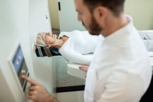
Dr Sherief Elsayed on Testing Yourself for Slipped Disc at Home

How accurate is the straight leg raise test at home?
The straight leg raise test has reasonable sensitivity for detecting nerve root irritation, but it’s not definitive. A positive test (leg pain radiating below the knee before 60 degrees) suggests nerve involvement, but clinical examination and imaging are needed for confirmation. False positives can occur with hamstring tightness or hip problems.
Can you have a slipped disc without leg pain?
Yes, disc herniations can cause isolated back pain without leg symptoms if the disc material doesn’t compress a nerve root. Some herniations are “contained,” meaning the outer disc layer remains intact. However, significant leg pain (sciatica) strongly suggests nerve compression and is more specific for disc herniation.
How long should I try home assessment before seeing a doctor?
If symptoms are mild and improving, observing for 1-2 weeks is reasonable. However, seek medical attention sooner if pain is severe, weakness develops, numbness spreads, or red flags appear. Don’t delay beyond 6 weeks if symptoms aren’t significantly improving with conservative measures.
Will a slipped disc heal on its own?
Many disc herniations improve naturally over 6-12 weeks as inflammation resolves and the body reabsorbs herniated material. Conservative treatment supports this natural healing process. However, some herniations require intervention if they cause progressive neurological problems or persistent disabling pain.
What's the difference between a bulging disc and a herniated disc?
A bulging disc shows outward extension of the disc margin beyond the vertebrae but the outer layer remains intact. A herniated (or prolapsed) disc means the inner material has broken through the outer layer. Herniations are more likely to compress nerves and cause symptoms, though both can be seen on MRI in people without symptoms.
Is it safe to exercise with a suspected slipped disc?
Gentle movement is generally beneficial and better than prolonged rest. Avoid exercises that worsen leg pain, especially those involving forward bending, heavy lifting, or twisting. Walking is usually well-tolerated. Once diagnosed, work with a physiotherapist to develop an appropriate exercise program that supports recovery without aggravating nerve compression.
A slipped disc medically known as disc herniation or disc prolapse occurs when the soft inner core of a spinal disc pushes through its tough outer layer, potentially compressing nearby nerves. Many people in Dubai and across the UAE experience sudden back or leg pain and wonder whether they might have a slipped disc, often before they can see a specialist.
While no home test can replace a proper medical examination and imaging, understanding basic self-assessment techniques can help you recognise warning signs and decide when to seek professional care. Dr. Sherief Elsayed, a senior UK-trained Consultant Spine Surgeon in Dubai, emphasises that self-awareness is valuable, but accurate diagnosis requires clinical expertise and often advanced imaging.
This guide explains how patients can perform simple observations at home, what symptoms matter most, and when self-testing should transition to urgent professional assessment.
What Is a Slipped Disc and Why Does It Matter?
A slipped disc refers to the displacement or herniation of one of the cushioning discs between the vertebrae in your spine. These discs act as shock absorbers, allowing flexibility and protecting the bones from rubbing together.
When a disc herniates, the inner gel-like material (nucleus pulposus) bulges or ruptures through the outer fibrous ring (annulus fibrosus). This can irritate or compress nearby spinal nerves, leading to:
- Local back pain at the level of the herniation
- Radiating leg pain (sciatica) if the disc affects lumbar nerve roots
- Numbness or tingling in specific areas of the leg or foot
- Muscle weakness in severe cases
- Bladder or bowel dysfunction in rare, emergency situations
In Dubai’s active lifestyle, whether lifting at the gym, long driving commutes, or desk-based work disc herniations are increasingly common. Recognising the symptoms early allows for timely, often conservative treatment that avoids surgery.
Can You Actually Test Yourself for a Slipped Disc at Home?
The short answer is: not definitively, but you can observe important clues.
Dr. Sherief Elsayed explains that while patients can identify patterns consistent with nerve compression, only clinical examination combined with MRI imaging can confirm disc herniation and its severity. However, certain movements and sensations provide meaningful information about whether nerve irritation is present.
What Home Observations Can Tell You
Home self-testing helps you:
- Recognise whether pain follows a nerve pattern (radicular pain)
- Identify positions that worsen or relieve symptoms
- Monitor progression of weakness or numbness
- Decide whether to seek urgent or routine assessment
What Home Testing Cannot Tell You
Self-assessment cannot:
- Confirm the exact location or size of herniation
- Differentiate disc herniation from other causes like piriformis syndrome or spinal stenosis
- Determine whether surgery is needed
- Rule out red flag conditions requiring emergency care
How Patients Usually Describe Slipped Disc Symptoms
Before exploring self-tests, it’s important to understand typical symptom patterns. Patients often describe:
- Sharp, shooting pain down one leg, worse than back pain itself
- Pain that worsens with sitting, bending forward, or coughing
- Relief when lying flat or walking gently
- Tingling or “pins and needles” in specific areas (e.g., outer calf, big toe)
- Feeling of weakness when standing on tiptoes or heels
- Sudden onset after lifting, twisting, or prolonged sitting
These patterns suggest nerve root involvement, which is the hallmark of disc herniation affecting lumbar nerves.
Simple Self-Assessment Techniques You Can Try at Home
While these observations don’t replace medical diagnosis, they help patients understand their symptoms better.
1. The Straight Leg Raise Test
What it assesses: Nerve root tension, particularly in L5 or S1 nerve roots commonly affected by lower lumbar disc herniations.
How to perform:
- Lie flat on your back on a firm surface
- Keep both legs straight
- Slowly raise the affected (painful) leg while keeping the knee straight
- Note at what angle pain begins or worsens
What it means:
- Positive test: Sharp leg pain (not just hamstring tightness) that shoots down the leg before reaching 60 degrees suggests nerve root irritation
- Negative test: Able to raise leg to 70+ degrees without radiating leg pain
Important notes:
- Hamstring tightness alone is not a positive test
- Pain should radiate below the knee to be significant
- Pain in the back only is less specific
2. The Slump Test (Seated Nerve Tension)
What it assesses: Nerve tension throughout the lower spine and leg.
How to perform:
- Sit on the edge of a chair or bed
- Slouch forward, rounding your lower back
- Tuck your chin to your chest
- Slowly straighten one knee, bringing your foot up
What it means:
- Positive test: Reproduces shooting leg pain or increases existing symptoms
- This suggests nerve involvement consistent with disc herniation
3. Toe and Heel Walking Test
What it assesses: Muscle strength controlled by specific nerve roots.
How to perform:
- Walk on your toes for 10-15 steps (tests S1 nerve root, calf strength)
- Walk on your heels with toes pointed up for 10-15 steps (tests L5 nerve root, ankle dorsiflexion)
What it means:
- Weakness on one side suggests nerve compression
- Inability to perform either test on the affected side is concerning
- Compare both sides asymmetry is more significant than bilateral difficulty
4. Dermatomal Sensation Check
What it assesses: Sensory changes following nerve distribution patterns.
How to perform:
- Gently touch or brush different areas of both legs
- Compare sensation between sides
- Key areas to check:
- Outer thigh (L2-L3)
- Front of knee (L4)
- Inner shin and big toe (L5)
- Outer foot and little toes (S1)
What it means:
- Numbness in specific patterns suggests which nerve root is affected
- Patchy or non-anatomical numbness is less specific
5. Movement Pattern Observation
What it assesses: Positions that increase or decrease nerve compression.
Observe:
- Pain worsens with: Sitting, bending forward, coughing, sneezing, bearing down
- Pain improves with: Lying flat, gentle walking, standing, extending backward
What it means:
- Worsening with forward bending and sitting suggests disc herniation
- This pattern reflects increased disc pressure in flexion
Dr. Sherief Elsayed’s Clinical Approach: Why Self-Testing Has Limits
While these home observations provide useful information, Dr. Sherief Elsayed emphasises several critical points that patients often overlook:
Physical Examination Goes Beyond Simple Tests
A comprehensive clinical examination includes:
- Detailed neurological assessment of reflexes, strength, and sensation
- Gait analysis to observe subtle asymmetries
- Palpation of the spine to identify tenderness or muscle spasm
- Range of motion testing in multiple directions
- Specialized provocative tests like Lasègue’s sign and femoral nerve stretch
Imaging Reveals What Examination Cannot
Even with a thorough clinical assessment, MRI imaging remains essential because it:
- Confirms the presence and exact location of disc herniation
- Shows the degree of nerve compression
- Identifies other pathology like spinal stenosis or tumours
- Guides treatment planning conservative versus surgical
Dr. Sherief Elsayed, a Spine Doctor in Dubai for herniated disc assessment, stresses that treating symptoms without understanding the underlying cause risks missing important diagnoses.
Not All Leg Pain Is a Slipped Disc
Many conditions mimic disc herniation:
- Piriformis syndrome: Muscle compression of the sciatic nerve
- Spinal stenosis: Narrowing of the spinal canal
- Sacroiliac joint dysfunction: Pelvic joint inflammation
- Hip pathology: Arthritis or labral tears
- Peripheral nerve entrapment: Compression outside the spine
Accurate diagnosis requires differentiating between these possibilities.
Real-World Scenarios Common in Dubai and the UAE
Understanding how daily activities contribute to disc herniation helps patients make better decisions.
Scenario 1: Office Worker with Prolonged Sitting
Profile: 35-year-old working 9+ hours daily at a desk, minimal breaks
Risk factors:
- Sustained forward flexion increases disc pressure
- Weak core muscles fail to support spine
- Prolonged sitting reduces disc nutrition
Self-testing application: Straight leg raise and slump test help identify nerve compression. If positive, consider ergonomic adjustments and standing breaks.
Scenario 2: Gym Enthusiast with Poor Lifting Technique
Profile: 28-year-old lifting heavy weights without proper form
Risk factors:
- Forward bending with load dramatically increases disc pressure
- Sudden twisting under load can cause acute herniation
- Inadequate warm-up and core bracing
Self-testing application: Weakness on toe or heel walking may indicate acute nerve compression requiring immediate medical attention.
Scenario 3: Long-Distance Driver
Profile: 42-year-old with daily commutes exceeding 2 hours
Risk factors:
- Vibration from driving increases disc stress
- Poor lumbar support in car seats
- Prolonged sitting in fixed position
Self-testing application: Worsening pain when sitting versus standing suggests disc involvement. Observation of symptom patterns helps guide treatment timing.
Scenario 4: Manual Labourer with Repetitive Bending
Profile: 50-year-old with years of repetitive forward bending and lifting
Risk factors:
- Cumulative disc degeneration over time
- Repetitive stress weakens disc structure
- Age-related loss of disc hydration
Self-testing application: Progressive weakness or numbness despite rest is concerning and warrants urgent assessment.
When Should You Worry and Seek Urgent Care?
While most disc herniations improve with conservative treatment, certain symptoms require immediate medical attention.
Red Flags Requiring Emergency Assessment
Seek urgent care if you experience:
- Bladder dysfunction: Inability to urinate, loss of bladder control, or lack of sensation when passing urine
- Bowel dysfunction: Loss of bowel control or inability to feel bowel movements
- Saddle anaesthesia: Numbness in the groin, inner thighs, or around the genitals and anus
- Progressive leg weakness: Rapidly worsening ability to walk or move the foot
- Severe pain following trauma: High-impact injury with severe, unrelenting pain
- Bilateral leg symptoms: Weakness, numbness, or pain in both legs simultaneously
These symptoms suggest cauda equina syndrome a surgical emergency requiring decompression within hours to prevent permanent damage. Dr. Sherief Elsayed has written extensively about this condition and when intervention becomes critical (When Is Sciatica Surgery Urgent?).
Urgent (Non-Emergency) Assessment Needed
Schedule prompt medical review if:
- Pain is severe and not improving after 48-72 hours
- You cannot bear weight or walk normally
- Numbness is spreading or worsening
- Weakness is developing in the foot or leg
- Conservative measures (rest, medication) provide no relief
Routine Assessment Appropriate
Many disc herniations improve naturally. Schedule a routine consultation if:
- Pain is manageable with over-the-counter medication
- Symptoms are gradually improving
- You can perform daily activities with minor discomfort
- No weakness or progressive numbness exists
What Happens After Self-Assessment?
If your home observations suggest nerve involvement, the next steps typically include:
Step 1: Clinical Consultation
A Spine Surgeon in Dubai will perform:
- Detailed history of symptom onset and progression
- Comprehensive neurological examination
- Assessment of functional limitations
- Discussion of lifestyle factors contributing to symptoms
Step 2: Imaging if Indicated
MRI scanning is typically ordered when:
- Symptoms persist beyond 6 weeks
- Red flags are present
- Weakness or progressive neurological deficit exists
- Surgery may be considered
Not every patient with back pain needs an MRI immediately. Many cases resolve with conservative care, and imaging is reserved for those who don’t improve or have concerning features.
Step 3: Treatment Planning
Treatment follows a stepwise approach:
- Conservative management (first-line for most patients):
- Activity modification
- Physical therapy and core strengthening
- Anti-inflammatory medications
- Short-term pain relief with appropriate medications
- Epidural injections if symptoms persist
- Minimally invasive options if conservative care fails:
- Transforaminal epidural injections
- Selective nerve root blocks
- Surgical intervention reserved for:
- Cauda equina syndrome
- Progressive neurological deficit
- Persistent, disabling pain after 12+ weeks of conservative care
- Severe nerve compression confirmed on imaging
Dr. Sherief Elsayed prioritises conservative treatment whenever appropriate, recognising that 80-90% of disc herniations improve without surgery.
Lifestyle Modifications That Support Recovery
While performing self-tests, patients can implement supportive measures:
Posture and Ergonomics
- Maintain neutral spine while sitting
- Use lumbar support in chairs and cars
- Take standing breaks every 30-45 minutes
- Adjust monitor height to avoid forward head posture
Activity Modification
- Avoid prolonged sitting initially
- Gentle walking encourages disc nutrition
- Avoid heavy lifting, especially with forward bending
- Gradually return to activities as pain permits
Core Strengthening
Once acute pain subsides:
- Focus on transverse abdominis activation
- Planks and bird-dog exercises
- Avoid crunches and sit-ups initially
- Progress under physiotherapy guidance
Sleep Positioning
- Side-lying with pillow between knees
- Avoid stomach sleeping
- Use appropriate mattress support
Common Misconceptions About Slipped Discs
“If I can move, it’s not a slipped disc”
Reality: Many people with confirmed herniations remain mobile. Severity varies widely depending on herniation size, location, and individual pain tolerance.
“An MRI will show everything wrong with my back”
Reality: MRI shows structure, not pain. Many people have disc herniations on imaging with no symptoms. Correlation between imaging and clinical findings is essential.
“Surgery is the only fix”
Reality: Surgery is rarely the first option. Most herniations respond to conservative care over weeks to months.
“I should rest in bed until it heals”
Reality: Prolonged bed rest weakens muscles and delays recovery. Gentle movement and activity modification are preferable.
Why Professional Assessment Remains Essential
Self-testing provides preliminary information but cannot replace expert evaluation. Dr. Sherief Elsayed’s approach integrates clinical findings, imaging, and patient goals to create personalised treatment plans.
Diagnosis Beyond Imaging
Pain is a symptom, not a diagnosis. Understanding the root cause whether mechanical stress, inflammatory processes, or neurological compromise guides effective treatment.
Treating the Person, Not the Scan
Two patients with identical MRI findings may require completely different treatments based on symptoms, lifestyle, and response to initial management.
Shared Decision-Making
Dr. Sherief Elsayed involves patients in treatment decisions, explaining options clearly and setting realistic expectations for recovery timelines and outcomes.
Conclusion
Testing yourself for a slipped disc at home can help you recognise patterns consistent with nerve compression and make informed decisions about seeking care. Simple observations like the straight leg raise test, toe and heel walking, or noting symptom patterns provide valuable preliminary information.
However, accurate diagnosis requires clinical examination and often MRI imaging. Self-assessment should never delay professional evaluation, especially when red flags like bladder dysfunction, progressive weakness, or saddle anaesthesia are present.
If you’re experiencing persistent back or leg pain, consult a qualified spine specialist who can perform a thorough assessment, order appropriate imaging, and develop a treatment plan tailored to your specific condition. Most disc herniations improve with conservative care, and surgery is reserved for those with specific indications or who don’t respond to non-operative treatment.
Early recognition combined with expert guidance offers the best chance for successful recovery and return to normal activities.
Table of Contents
Recent Articles

Why Leaning Backwards Makes Your Back Pain Worse and What It Means for Your Spine
Why Leaning Backwards Makes Your Back Pain Worse and What It Means for Your Spine Is facet joint pain the same as arthritis? Facet joint degeneration is essentially osteoarthritis of
Why an MRI Report Alone Is Not Enough to Decide on Spine Treatment in the UAE
Why an MRI Report Alone Is Not Enough to Decide on Spine Treatment in the UAE I have a disc protrusion on my MRI but no leg pain. Do I

General Anaesthesia vs Sedation, What Every Patient in Dubai Should Know Before Surgery
General Anaesthesia vs Sedation, What Every Patient in Dubai Should Know Before Surgery Will I feel anything during general anaesthesia? No. General anaesthesia eliminates consciousness, sensation, and awareness. You will