
Why Hip Disease and Sciatica Feel So Similar, A UAE Surgeon Explains
How is avascular necrosis diagnosed?
AVN is best diagnosed on MRI, which detects the characteristic signal changes of bone infarction before any structural collapse is visible on X-ray. In advanced cases as described here, plain X-ray is sufficient to show the diagnosis. Staging is based on the degree of femoral head involvement and collapse.
Can AVN be treated without surgery?
In early stages (before femoral head collapse), conservative measures, reduction of risk factors such as steroids, and in selected cases, core decompression (a surgical procedure to reduce pressure within the femoral head and restore blood flow) may slow progression. Once significant collapse has occurred, joint replacement is typically the only effective treatment.
Can the same condition affect both hips?
Yes. Bilateral AVN is not uncommon, particularly in patients with systemic causes such as steroid use, sickle cell disease, or autoimmune conditions. Both hips should be assessed even when only one is symptomatic.
How do I know if my groin pain is from the hip or the spine?
The key distinguishing features are: hip pain is typically groin-to-anterior-knee, worsened by weight-bearing and rotation of the hip, and associated with restricted hip movement on examination. Spinal nerve root pain typically travels below the knee in a specific dermatomal pattern and is associated with neurological signs. A clinical assessment examining both structures is the definitive way to distinguish them.
Why did six doctors miss this diagnosis?
Most commonly, the hip is not examined when a patient presents with leg pain in a spine-focused consultation. Additionally, coincidental degenerative spine changes on imaging can be incorrectly attributed as the pain source without confirming clinical correlation. Systematic examination of all relevant anatomical structures prevents this error.
What is the recovery like after total hip replacement?
Most patients are walking within 24 hours of surgery and discharged within two to three days. Return to normal daily activity takes four to six weeks. Return to unrestricted activity including sport takes three to six months. Pain relief is typically dramatic and immediate compared to the pre-operative level.
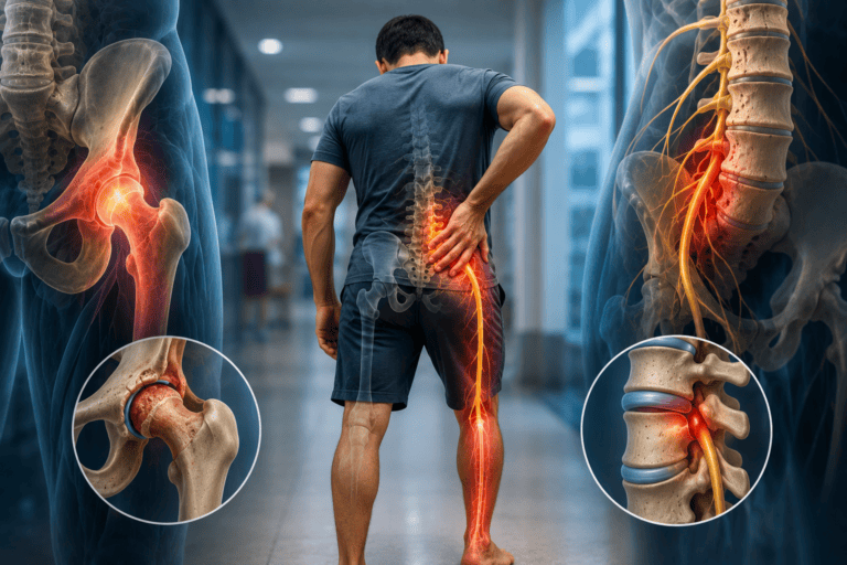
Six doctors over eight months. Severe leg pain. No diagnosis. This is not an unusual story in spine and orthopaedic medicine, and it illustrates one of the most persistent diagnostic challenges in musculoskeletal care: the overlap between hip pathology and lumbar spine pathology in terms of where pain is felt and how it presents.
Dr. Sherief Elsayed, Consultant Spine Surgeon in Dubai, describes a 60-year-old patient whose correct diagnosis was reached within a single consultation, not by ordering more investigations, but by examining the right structure.
The Clinical Case: Six Doctors, Eight Months, No Answer
Dr. Sherief Elsayed describes the encounter directly: “Around two weeks ago, I saw a 60-year-old gentleman who was complaining of severe leg pain. Over the previous eight months, he’d seen six different doctors, none of whom could quite figure out where his pain was coming from.”
The first clinical observation was made before the patient said a word. “As soon as he stepped into my room, I noted that he had difficulty walking. He was taking his foot off his leg as quickly as he could.”
This antalgic gait pattern. The tendency to bear weight on the affected limb for as short a time as possible, is a non-specific but important sign. It tells the clinician that weight-bearing is painful and that the patient is making every movement count to minimise the time spent loading whatever structure is producing the pain.
The history then provided the key localising detail: “He told me that he had a lot of groin pain, which went along the front of his thigh down to his knee.”
Why This Distribution Is Not Sciatica
The clinical reasoning at this point is exactly the same as that described in the published article How a Spine Doctor in Dubai Tells the Difference Between Sciatica and Hip Arthritis. Groin pain that travels down the front of the thigh to the knee does not follow the sciatic nerve path, which runs from the lower back through the buttock and down the back of the leg.
Dr. Sherief Elsayed notes: “That’s not very common for sciatica.”
True sciatica travels below the knee into the calf, ankle, or foot. It originates from the lower lumbar nerve roots (typically L4, L5, or S1) and follows specific dermatomal patterns that are well-mapped and recognisable. Pain confined to the groin and anterior thigh, stopping at or above the knee, suggests one of several other anatomical sources:
- The hip joint, which refers pain into the groin and anterior thigh as its primary pattern
- The upper lumbar nerve roots (L2 or L3), which is less common but possible
- The femoral nerve, producing femoral neuralgia with anterior thigh pain and reduced knee jerk
- The sacroiliac joint, which can refer into the buttock and posterior thigh
In a 60-year-old man with groin-to-anterior-knee pain and antalgic gait, the hip is the first structure to examine, not the lumbar spine.
The Examination That Made the Diagnosis
“When I examined him, I noted that he had severe restriction of his hip movements, and that led me to believe that it was his hip that was the cause of his leg pain, not sciatica coming from his lower back.”
Hip examination is the decisive step. The hip joint is assessed through its range of motion in all planes, flexion, extension, abduction, adduction, internal and external rotation. A healthy hip moves through a large and comfortable range. A diseased hip, whether from osteoarthritis, avascular necrosis, or other pathology, is restricted, painful at the end of range, and produces characteristic pain reproduction with specific movements.
Internal rotation in particular is typically the first movement to be lost in hip joint disease. When the examiner rotates the hip internally (turning the foot outward while the hip is in a neutral position), a patient with hip pathology will experience significant pain and resistance well before the full range is reached.
Severe restriction of hip movements, as Dr. Sherief Elsayed found, is clinically incompatible with a normal hip joint. The lumbar spine examination, by contrast, would be expected to show minimal restriction in a patient whose pain is originating entirely from the hip.
The principle here applies broadly: a positive hip examination in the presence of anterior thigh pain points strongly to the hip. A positive spinal examination, reduced straight leg raise, neurological deficit in a dermatomal pattern, lumbar spine tenderness, points to the spine. When the hip examination is the positive finding, investigate the hip.
The Diagnosis: Avascular Necrosis
“I x-rayed his hip, and lo and behold, he has a severe disease of his hip called avascular necrosis, where the head of the hip breaks down and causes excruciating pain. Only possible treatment for this at this late stage is a total hip replacement, and I’ve sent him on to one of my excellent hip colleagues for that to be done.”
Avascular necrosis (AVN) of the femoral head, also called osteonecrosis, is a condition in which the blood supply to the femoral head is disrupted, causing the bone to die and the head of the hip to gradually collapse and fragment. Unlike osteoarthritis, which is a degenerative process, AVN involves infarction of the bone itself.
Causes of avascular necrosis of the femoral head include:
- Prolonged corticosteroid use (the most common cause in younger patients)
- Excessive alcohol consumption
- Previous hip trauma or dislocation
- Sickle cell disease and other haemoglobinopathies
- Systemic lupus erythematosus and other connective tissue disorders
- Decompression sickness (in divers)
- In some cases, no identifiable cause (idiopathic AVN)
Why AVN produces such severe pain:
As the femoral head collapses, the normally smooth, congruent ball-and-socket joint becomes irregular and incongruent. Every movement grinds irregular bony surfaces against each other. The pain is not simply arthritic. It has a quality of mechanical grinding combined with the deep bony ache of ischaemic bone. Patients often describe it as one of the most severe musculoskeletal pains they have experienced.
Why the hip collapse was already severe:
AVN progresses through stages. In the early stages, the bone changes are detectable only on MRI. As the condition advances, the femoral head collapses and the joint space narrows. At the stage this patient presented, the X-ray was sufficient to show the diagnosis, meaning the structural damage was well advanced. Eight months of misdiagnosis had allowed the condition to progress without the specific treatment that might have slowed it or altered the surgical approach.
Why Was This Misdiagnosed for Eight Months?
The patient saw six doctors without a correct diagnosis. There are several reasons why this pattern occurs.
Failure to examine the hip: In a patient presenting with leg pain, particularly in a spine-focused environment, the lumbar spine is often investigated before the hip is examined. If imaging of the lumbar spine shows degeneration (which it almost certainly will in a 60-year-old), the degeneration may be accepted as the explanation without a full hip examination being performed.
Reliance on imaging over examination: An MRI of the lumbar spine may show disc changes, mild foraminal narrowing, or facet joint degeneration, all common findings at this age, which can be incorrectly attributed as the pain source without the clinical correlation that confirms or refutes the attribution.
The overlap in symptom location: Groin pain and anterior thigh pain are genuinely shared territory between hip pathology and upper lumbar pathology. Without a systematic approach to differential diagnosis that includes specific hip examination, the distinction is not made.
Patient description: Patients commonly describe their pain as “my hip” when pointing to their lateral thigh, which is actually the greater trochanteric region and often related to trochanteric bursitis rather than hip joint pathology. This imprecision in lay anatomical language can mislead clinicians who do not ask follow-up questions about the exact location and distribution.
The Lesson: Examine the Structure the Pain Is Pointing To
Dr. Sherief Elsayed’s approach in this case was direct and correct: groin-to-knee pain in a 60-year-old with antalgic gait pointed to the hip, the hip was examined, the examination was positive, and the hip was imaged. The diagnosis was made in a single consultation.
This is the clinical sequence that eight months and six doctors had not followed. Each step depends on the preceding one, and the sequence must start with examining the right structure. A Spine Doctor in Dubai who applies systematic anatomical reasoning to every lower limb pain presentation will examine both the spine and the hip in any case where the distribution of pain is ambiguous, precisely because the overlap is real and the consequences of misdiagnosis can be prolonged suffering and unnecessary investigation.
The complementary article on this topic, Pain in the Front of Your Leg Is Not Always Sciatica, explores the full range of conditions that produce anterior thigh pain and how each is distinguished on clinical assessment.
UAE-Specific Considerations
In the UAE, several factors increase the likelihood of AVN presenting in the clinical population.
Corticosteroid use: Steroids are widely prescribed for a range of conditions in the UAE, including respiratory disease, inflammatory conditions, and skin disorders. Prolonged high-dose steroid use is the leading cause of non-traumatic AVN, and any patient who has received substantial steroid courses should be aware of this risk.
Haemoglobinopathies: Sickle cell disease and sickle cell trait are prevalent in certain communities represented in the UAE’s diverse population, particularly from sub-Saharan Africa and parts of the Arab world. Sickle cell crises cause vascular occlusion that can affect the femoral head.
Alcohol: While alcohol consumption is lower in the Muslim-majority population of the UAE than in many Western countries, it is not absent among the large expatriate population, and alcohol-related AVN is a recognised clinical entity.
Expert Summary
Eight months of misdiagnosis ended with a single consultation that included a hip examination. The pain distribution, groin to anterior knee, was not consistent with classic sciatica. The antalgic gait was visible on entry. The restricted hip movements confirmed the joint as the pain source. The X-ray confirmed severe AVN requiring total hip replacement.
The lesson is that the correct diagnosis of lower limb pain requires examining both the spine and the hip. When the spine examination is unremarkable and the hip examination is severely restricted, the hip is the problem regardless of what the lumbar MRI shows. To avoid the pattern of prolonged misdiagnosis this patient experienced, a first assessment with a Consultant Spine Surgeon in Duba who systematically examines all relevant structures is the most direct route to an accurate diagnosis and the right treatment.
Table of Contents
Recent Articles
How Dr. Sherief Elsayed Spots Spinal Tumours Before Paralysis Sets In
How Dr. Sherief Elsayed Spots Spinal Tumours Before Paralysis Sets In If I have no symptoms, why would a tumour on my spine need urgent treatment? The absence of symptoms
Why a Custom 3D Scanned Brace Works Better for Scoliosis in Dubai
Why a Custom 3D Scanned Brace Works Better for Scoliosis in Dubai Is 3D scanning for scoliosis braces widely available in Dubai? 3D scanning for brace manufacture is available at
What Happens When a Spine Cage Is Placed in the Wrong Position?
What Happens When a Spine Cage Is Placed in the Wrong Position? How is cage malpositioning diagnosed? Post-operative imaging is essential. Plain X-rays can identify obvious cage malpositioning. CT scanning