
How Dr. Sherief Elsayed Spots Spinal Tumours Before Paralysis Sets In
If I have no symptoms, why would a tumour on my spine need urgent treatment?
The absence of symptoms reflects the spinal cord’s current capacity to tolerate compression, not the absence of risk. A tumour that continues to grow will eventually exceed the cord’s tolerance, at which point neurological decline can occur rapidly. Treating the tumour while the patient remains neurologically intact gives the best chance of preserving normal function.
How is a spinal tumour diagnosed if there are no symptoms?
Tumours without neurological symptoms are typically found incidentally on imaging performed for another reason, through surveillance imaging in patients with a known cancer history, or through investigation of early, non-specific symptoms such as localised back pain that prompts an MRI scan.
What is the difference between a primary spinal tumour and a metastatic spinal tumour?
A primary spinal tumour originates within the spine or its coverings. A metastatic spinal tumour has spread to the spine from a cancer elsewhere in the body, most commonly the lung, breast, prostate, kidney, or thyroid. The distinction affects treatment planning, as metastatic disease is generally managed with a combination of surgery, radiotherapy, and systemic oncological treatment, while primary spinal tumours may be curable with appropriate surgical resection depending on the specific type.
Can a spinal tumour be removed completely?
This depends on the tumour type, location, and relationship to critical structures including the spinal cord and major blood vessels. Some primary tumours can be resected completely with curative intent. Metastatic tumours are more often treated with decompression and stabilisation rather than complete removal, as the treatment goal is usually to preserve function and quality of life alongside broader oncological management.
What is the recovery like after surgery for a spinal tumour with no pre-operative symptoms?
Patients who undergo surgery while neurologically intact generally have the best post-operative outcomes, as the goal of surgery is to preserve existing function rather than restore lost function. Recovery depends on the extent of surgery, whether instrumentation and fusion were required, and any adjuvant treatment planned, but the neurological prognosis in this scenario is considerably more favourable than in patients who present after paralysis has already developed.
Should I ask for spinal imaging if I have a history of cancer, even without back pain?
Discuss this directly with your oncology team, as surveillance imaging protocols vary depending on cancer type and stage. Patients with cancers known to commonly metastasise to bone, including lung, breast, prostate, kidney, and thyroid cancer, should have a low threshold for spinal imaging if any new back pain or neurological symptoms develop, even if these seem mild or non-specific.
Some of the most consequential decisions in spine surgery are made not when a patient is already paralysed, but in the narrow window before that happens, when a tumour is visibly present, actively growing, and neurologically silent. A patient who currently has no weakness and no bladder or bowel symptoms can look, from their own perspective, entirely well. The imaging tells a different story, and the gap between what the patient feels and what the scan shows is precisely where a spine surgeon’s judgement matters most.
Dr. Sherief Elsayed, Consultant Spine Surgeon in Dubai, walks through exactly this kind of case, describing what a spinal tumour looks like on imaging before it has caused any clinical symptoms, and explaining why the absence of symptoms today does not mean the absence of urgency.
The Case: A Tumour With No Symptoms Yet
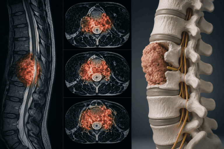
Dr. Sherief Elsayed describes the imaging: “This is a really interesting case. This is a patient with what looks like a spine tumour. You can see here what the normal vertebra looks like, what a normal disc looks like. There’s the spinal cord at the back. Now look at this vertebra. It’s full of tumour material and it’s coming outside the bone and around the spinal cord.”
The description continues with a vivid illustration of the three-dimensional extent of the tumour: “If we chop the patient up, salami slices, you can see here, all this is tumour. This is the spinal cord coming out of the screen. The tumour is going all around the spinal cord.”
This “salami slice” description refers to viewing sequential axial (cross-sectional) MRI images, each one representing a thin slice through the patient’s anatomy at a specific level, much as slicing through a loaf of bread reveals the internal structure at each point along its length. Reviewing these sequential slices allows the surgeon to build a complete three-dimensional understanding of exactly how much of the vertebra is replaced by tumour and how far that tumour extends around the circumference of the spinal cord.
Why “No Weakness, No Bladder or Bowel Problems” Is Not Reassuring
The clinical status of the patient at this point in the case is genuinely important: “At the moment, he has no weakness. He has no problems with his bladder or bowel.”
For many medical conditions, the absence of symptoms would be a reassuring finding, suggesting either that the pathology is not significant or that it can be safely monitored. For a spinal tumour that is circumferentially surrounding the spinal cord, the absence of symptoms carries a very different meaning.
Why the spinal cord can be surrounded by tumour without symptoms:
The spinal cord has some capacity to tolerate slowly progressive compression before neurological function is lost. As a tumour gradually expands and encroaches on the space around the cord, the cord itself may be displaced and compressed to a degree without immediately losing function, particularly if the growth is slow enough to allow some degree of physiological adaptation.
This tolerance has limits, and importantly, those limits are not predictable in advance for an individual patient. The cord’s compensatory capacity can appear robust right up until a threshold is crossed, at which point deterioration can be sudden rather than gradual. A patient can have a tumour circumferentially surrounding their cord one week with completely normal neurological examination, and present the following week with rapidly progressive weakness once the compression exceeds what the cord could continue to tolerate.
The Prediction: “If We Leave This…”
Dr. Sherief Elsayed states the trajectory plainly: “But if we leave this, that tumour is going to continue to grow. It’s going to crush his spinal cord. And there’s a significant chance of paralysis.”
This is not a hypothetical caution offered defensively. It is a specific clinical prediction based on the biology of untreated tumour growth and the anatomical reality of a mass that already surrounds the spinal cord with no room remaining to expand without directly compressing neural tissue further.
The clinical reasoning behind acting before symptoms appear:
As established in the companion article How Long Can a Compressed Nerve Wait Before Surgery, the longer neural tissue is compressed, the less likely it is to recover after decompression. This principle applies with particular force to spinal cord compression from tumour.
If treatment is delayed until the patient develops weakness, the surgical decompression that follows is addressing established, ongoing cord compression that has already begun causing measurable neurological injury. The outcome in this scenario is less predictable and often incomplete, because some of the neurological damage may already be irreversible by the time surgery is performed.
If treatment is undertaken while the patient remains neurologically intact, the surgery removes the compressive tumour before the cord has been subjected to the sustained pressure that produces lasting injury. The neurological outcome in this scenario is substantially better, because the surgery is preventive rather than rescue.
This is precisely why Dr. Sherief Elsayed’s assessment of “a significant chance of paralysis” if the tumour is left untreated is not an overstatement designed to prompt action. It is an accurate representation of the trajectory that circumferential spinal cord compression from a growing tumour follows if surgery is delayed until symptoms force the issue.
What Determines the Urgency and the Surgical Approach
Several factors inform how a spine surgeon assesses the urgency of intervention in a case like this. A Spinal Cord Surgeon in Dubai weighs canal compromise, growth rate, and tumour biology together when planning treatment. of intervention in a case like this.
Degree of canal compromise: The percentage of the spinal canal occupied by tumour, and the degree to which the cord itself is compressed and displaced from its normal position, are directly measurable on MRI and provide an objective assessment of how much reserve space remains before further growth produces critical compression.
Rate of growth: Where prior imaging is available for comparison, the rate of tumour growth over time provides important information about how quickly the situation is likely to change. A tumour that has grown rapidly over a short interval suggests a shorter window before symptoms develop than one that has been stable or slowly progressive over a longer period.
Tumour type and biology: Different tumour types, whether primary spinal tumours or metastatic deposits from a cancer elsewhere in the body, have different growth rates and different treatment approaches. Biopsy or histological diagnosis, where safely obtainable, informs whether surgery, radiotherapy, or a combination is the most appropriate primary treatment.
Patient factors: The patient’s overall fitness for surgery, their other medical conditions, and in the case of metastatic disease, their broader oncological prognosis, all factor into the urgency and extent of intervention that is appropriate. A Spine Tumour Surgeon in Dubai will weigh all of these factors together in formulating a recommendation, rather than applying a single fixed rule to every tumour case.
Why Early Detection Matters So Much
This case illustrates why early detection of spinal tumours, before any neurological symptoms develop, is genuinely valuable rather than an incidental benefit of unrelated imaging.
How such tumours are often found:
Some spinal tumours are detected incidentally, identified on imaging performed for an unrelated reason, such as back pain that prompted an MRI that then revealed the mass. Others are found through surveillance imaging in patients with a known history of cancer elsewhere in the body, where regular monitoring for metastatic spread is part of routine oncological care. Some present with early, non-specific symptoms such as localised back pain, without any neurological features, that prompt imaging before more serious symptoms develop.
Why any of these pathways represents an opportunity:
Whatever the reason imaging was obtained, finding a significant spinal tumour before it has produced neurological compromise represents a genuine clinical opportunity. It allows the surgical and oncological team to plan treatment methodically, arrange any necessary additional investigations, and undertake surgery under controlled, elective circumstances rather than as an emergency response to sudden paralysis.
The published article Why Lung Cancer Can Spread to the Spine, Dr. Sherief Elsayed Explains covers a related scenario where the tumour was found only after neurological symptoms had already developed, illustrating the less favourable circumstances that arise when detection occurs later in the disease course. The contrast between these two cases underscores the clinical value of vigilance and appropriate imaging thresholds in patients with risk factors or early, non-specific symptoms.
What Happens Next for This Patient
Following the identification of a tumour circumferentially surrounding the spinal cord in a neurologically intact patient, the appropriate next steps typically include further staging investigations (to determine whether the lesion is a primary spinal tumour or a metastasis from elsewhere), multidisciplinary discussion involving oncology, radiology, and spinal surgery, and planning for surgical decompression and stabilisation, with or without adjuvant treatment such as radiotherapy, depending on the specific tumour type identified.
The goal of surgery in this scenario is to remove or debulk the tumour sufficiently to decompress the spinal cord, reconstruct and stabilise the spine as needed (using techniques including bone cement and instrumentation as covered in the earlier articles in this series), and preserve the patient’s currently normal neurological function rather than attempting to restore function that has already been lost.
Expert Summary
A spinal tumour that circumferentially surrounds the spinal cord without yet producing weakness or bladder or bowel symptoms represents a genuine clinical urgency, not a reassuring finding. The cord’s capacity to tolerate compression without symptoms has limits that are not predictable for an individual patient, and once those limits are exceeded, neurological decline can be sudden and difficult to fully reverse. Dr. Sherief Elsayed’s assessment that untreated growth carries a significant chance of paralysis reflects the established biology of spinal cord compression, and it is precisely this understanding that drives the decision to intervene before symptoms force the issue, when outcomes are most favourable.
Table of Contents
Recent Articles
How Dr. Sherief Elsayed Spots Spinal Tumours Before Paralysis Sets In
How Dr. Sherief Elsayed Spots Spinal Tumours Before Paralysis Sets In If I have no symptoms, why would a tumour on my spine need urgent treatment? The absence of symptoms
Why a Custom 3D Scanned Brace Works Better for Scoliosis in Dubai
Why a Custom 3D Scanned Brace Works Better for Scoliosis in Dubai Is 3D scanning for scoliosis braces widely available in Dubai? 3D scanning for brace manufacture is available at
What Happens When a Spine Cage Is Placed in the Wrong Position?
What Happens When a Spine Cage Is Placed in the Wrong Position? How is cage malpositioning diagnosed? Post-operative imaging is essential. Plain X-rays can identify obvious cage malpositioning. CT scanning